
Atlas of Human Anatomy in Cross Section: Section 1. Head and Neck
Ronald A. Bergman, Ph.D., Adel K. Afifi, M.D., Jean J. Jew, M.D., and Paul
C. Reimann, B.S.
Peer Review Status: Externally Peer Reviewed
|
Upper Left Quadrant |
Lower Left Quadrant |
Lower Right Quadrant |
Upper Right Quadrant |
|
1. Superior frontal gyrus |
4. Cingulum |
8. Superior sagittal sinus |
13. Superficial temporal a. |
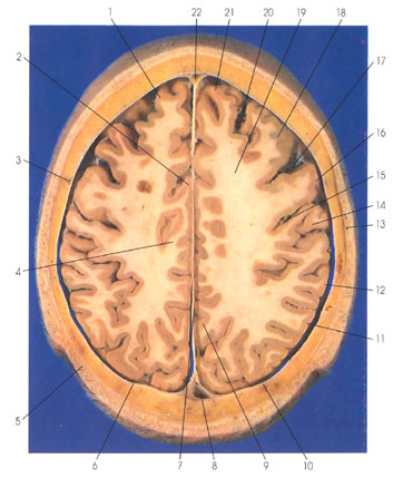
This section (looking down) is through the centrum semiovale (19) and superficial to the cerebral ventricles. The centrum semiovale (19) comprises the central white matter core of the cerebral hemispheres. The two hemispheres are separated by the falx cerebri (7), a dural fold within the interhemispheric fissure. The superior sagittal sinus is seen rostrally (22) and caudally (8) within the falx cerebri. The falx cerebri (7) is continuous with the dura mater around the cerebral hemispheres (10). Superficial to the dura mater (10) between it and the calvarium is the middle meningeal artery (3). Rupture of the middle meningeal artery, as occurs in some skull fractures, may lead to a life threatening epidural arterial hemorrhage, epidural hematoma. The superficial temporal artery ( 13) is seen within the scalp (5). In the frontal lobe are seen the superior (1, 21) and the middle (18) frontal gyri separated by the superior frontal sulcus (20). The precentral gyrus ( 16) is seen between the precentral (17) and the central (rolandic) (15) sulci. The precentral gyrus is the primary motor cortex. The central (rolandic) sulcus (15) separates the precentral gyrus (16) of the frontal lobe from the postcentral gyrus (14) of the parietal lobe. The postcentral gyrus (14) is the primary somatosensory (somesthetic) cortex. In the parietal lobe are also seen the supramarginal (12) and angular (11) gyri, two important gyri in association and language functions of the brain. On the medial surface of the parietal lobe is the precuneus gyrus (9). The cingulate gyrus (2) is seen on the medial surface of the hemisphere; its white core, the cingulum (4), is one of the important long association bundles of the cerebral hemispheres.
Next Page | Previous Page | Section Top | Title Page
Please send us comments by filling out our Comment Form.
Anatomy Atlases is licensed under a Creative Commons Attribution-NonCommercial-ShareAlike 4.0 International License. 
"Anatomy Atlases", the Anatomy Atlases logo, and "A digital library of anatomy information" are all Trademarks of Michael P. D'Alessandro, M.D.
Anatomy Atlases is funded in whole by Michael P. D'Alessandro, M.D. Advertising is not accepted.
Your personal information remains confidential and is not sold, leased, or given to any third party be they reliable or not.
The information contained in Anatomy Atlases is not a substitute for the medical care and advice of your physician. There may be variations in treatment that your physician may recommend based on individual facts and circumstances.
URL: http://www.anatomyatlases.org/