
Atlas of Human Anatomy in Cross Section: Section 1. Head and Neck
Ronald A. Bergman, Ph.D., Adel K. Afifi, M.D., Jean J. Jew, M.D., and Paul
C. Reimann, B.S.
Peer Review Status: Externally Peer Reviewed
|
Upper Left Quadrant |
Lower Left Quadrant |
Lower Right Quadrant |
Upper Right Quadrant |
|
1. Lamina terminalis |
11. Fimbria of Fornix |
20. Vermis of cerebellum |
28. Middle temporal gyrus |
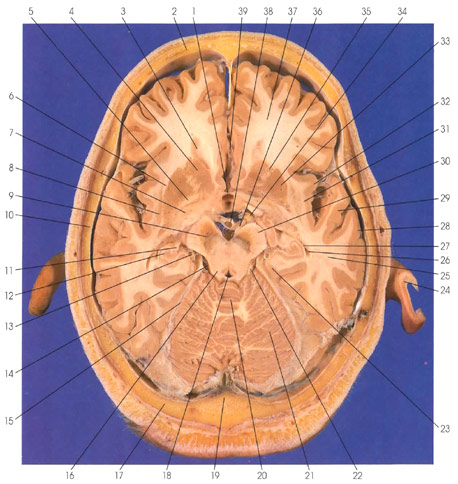
This is a section (looking down) through the basal ganglia, midbrain, and cerebellum. In the interhemispheric fissure rostrally is the anterior cerebral artery (39). On each side of the interhemispheric fissure are the frontal lobes (37). The floor of the frontal (anterior) horn of the lateral ventricle (3) is seen. The lateral ventricles on both sides are seen for the last time. The head of the caudate nucleus (4) lines the lateral wall of the frontal (anterior) horn of the lateral ventricle (3). The anterior limb of the internal capsule (35) separates the head of the caudate nucleus (4) from the putamen (33). The anterior commissure (5) is in close proximity to the putamen (33). Caudal to the anterior commissure (5) is the anterior perforated substance (7). Caudal to the interhemispheric fissure and in close proximity to the anterior commissure (5) is the lamina terminalis (1), landmark for the most rostral part of the embryonic neural tube. Lateral to the putamen (33) is the external capsule (6). The claustrum (8) is sandwiched between the external (6) and extreme (31) capsules. The midbrain is seen rostral to the cerebellum (20,21). Within the midbrain, the following structures are seen: aqueduct of Sylvius (18), superior colliculus (16), substantia nigra (9), and cerebral peduncle (30). The oculomotor nerve (36) is seen exiting from the inferior surface of the midbrain. In the subarachnoid space around the midbrain, the following arteries are seen: basilar (38), posterior communicating (34), posterior cerebral (13), and superior cerebellar (22). The ambient cistern (14) is seen in relation to the midbrain. Caudal to the midbrain is the cerebellum with its midline vermis (20) and lateral hemispheres (21). The tentorium cerebelli (15) is a dural fold between the cerebellum and occipital lobe. Caudal to the cerebellum is the occipital bone (17) and internal occipital crest (19). The temporal (inferior) horn of the lateral ventricle (26) makes its last appearance. The hippocampus (12) protrudes into the temporal (inferior) horn of the lateral ventricle (26). The alveus (27), an outflow tract from the hippocampus, is continuous with the fimbria of the fornix (11). The hippocampus (12) is seen as an invagination of the parahippocampal gyrus (23) into the temporal (inferior) horn of the lateral ventricle (26). The insula (island of Reil) (32) is seen in the depth of the lateral (sylvian) fissure. In the temporal lobe (25), the superior (29) and middle (28) temporal gyri are seen. The frontal (2), temporal (10), and occipital (17) bones and the auricle (24) are seen.
Next Page | Previous Page | Section Top | Title Page
Please send us comments by filling out our Comment Form.
Anatomy Atlases is licensed under a Creative Commons Attribution-NonCommercial-ShareAlike 4.0 International License. 
"Anatomy Atlases", the Anatomy Atlases logo, and "A digital library of anatomy information" are all Trademarks of Michael P. D'Alessandro, M.D.
Anatomy Atlases is funded in whole by Michael P. D'Alessandro, M.D. Advertising is not accepted.
Your personal information remains confidential and is not sold, leased, or given to any third party be they reliable or not.
The information contained in Anatomy Atlases is not a substitute for the medical care and advice of your physician. There may be variations in treatment that your physician may recommend based on individual facts and circumstances.
URL: http://www.anatomyatlases.org/