
Atlas of Human Anatomy in Cross Section: Section 1. Head and Neck
Ronald A. Bergman, Ph.D., Adel K. Afifi, M.D., Jean J. Jew, M.D., and Paul
C. Reimann, B.S.
Peer Review Status: Externally Peer Reviewed
|
Upper Left Quadrant |
Lower Left Quadrant |
Lower Right Quadrant |
Upper Right Quadrant |
|
1. Gyrus rectus of frontal lobe |
10. Semicircular canals |
15. Fourth ventricle |
21. Facial nerve |
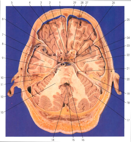
This section is through the pons (9, 16) and cerebellum (14, 17). The frontal and temporal lobes are seen. Within the calvarium, the frontal sinus (27) is seen. The lesser wing of the spheroid bone (6) and the petrous portion of the temporal bone (19) are seen. Within the petrous portion of the temporal bone (19), the mastoid air cells (11) and the semicircular canals (10) are seen. Within the dura mater, the sigmoid venous sinus (12) is seen. In the frontal lobe, the orbital gyri (4) are lateral and the gyrus rectus (1) is medial. The longitudinal (interhemispheric) fissure (29) separates the two frontal lobes. The middle cerebral artery (24) is seen within the lateral (sylvian) fissure. The amygdaloid nucleus (22) is seen within the temporal lobe (8). The anterior (26) and posterior (23) petroclinoid ligaments are seen. The internal carotid artery (3, 25) is seen within the cavernous sinus (25) and in close proximity to the optic nerve (28). The oculomotor nerve (5) is seen within the cavernous sinus (25). The tuber cinereum (2) is seen between the internal carotid arteries (3). The pons (9, 16) is connected to the cerebellum (14, 17) via the middle cerebellar peduncle (brachium pontis) (13). Within the pons, the tegmentum (16) is dorsal and the basis pontis (9) is ventral. The basilar artery (7) is ventral to the pons. The facial nerve (21) is seen exiting from the lateral surface of the pons. Within the cerebellum, the midline vermis (14) and the lateral cerebellar hemispheres (17) are seen. The fourth ventricle (15) lies between the vermis of the cerebellum (14) and the tegmentum of the pons (16). The tentorium cerebelli (18) is a dural fold between the cerebellum and occipital lobe. Outside the skull, the auricle (20) is seen.
Next Page | Previous Page | Section Top | Title Page
Please send us comments by filling out our Comment Form.
Anatomy Atlases is licensed under a Creative Commons Attribution-NonCommercial-ShareAlike 4.0 International License. 
"Anatomy Atlases", the Anatomy Atlases logo, and "A digital library of anatomy information" are all Trademarks of Michael P. D'Alessandro, M.D.
Anatomy Atlases is funded in whole by Michael P. D'Alessandro, M.D. Advertising is not accepted.
Your personal information remains confidential and is not sold, leased, or given to any third party be they reliable or not.
The information contained in Anatomy Atlases is not a substitute for the medical care and advice of your physician. There may be variations in treatment that your physician may recommend based on individual facts and circumstances.
URL: http://www.anatomyatlases.org/