
Atlas of Human Anatomy in Cross Section: Section 1. Head and Neck
Ronald A. Bergman, Ph.D., Adel K. Afifi, M.D., Jean J. Jew, M.D., and Paul
C. Reimann, B.S.
Peer Review Status: Externally Peer Reviewed
|
Upper Left Quadrant |
Lower Left Quadrant |
Lower Right Quadrant |
Upper Right Quadrant |
|
1. Sphenoid sinus |
7. Basilar a. |
13. Internal occipital crest |
20. Facial nerve |
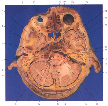
This is a section through the temporal lobe (22), cerebellum (15), and medulla oblongata (11, 12). Between the medulla oblongata(11, 12) and cerebellum (15) is the fourth ventricle (14). Within the medulla oblongata, the medullary pyramids (11) are ventrally placed and the medullary olives (12) are dorsal to the pyramids (11). Ventral to the medullary pyramids (11) is the abducens nerve (17), which exits the brain stem at the medulla pons junction. The ventral cochlear nucleus (16) is seen in a dorsolateral position in the medulla. The vestibulocochlear nerve ( 19) enters the medullary pontine junction dorsolaterally. The facial nerve (20) travels in close proximity to the vestibulocochlear nerve (19). Both are seen entering the internal auditory meatus (21). Ventral to the medullary pyramids (11) is the basilar artery (7) lying on the basiocciput (clivus) (27). The vertebral artery (10) is lateral to the medulla. The white matter core of the cerebellum is the corpus medullare of the cerebellum (15). Rostrally in the section are seen the nasal conchae (28). Caudal to the nasal cavity is the spheroid sinus (1). Within the orbital cavity two extraocular muscles are seen: the medial (26) and lateral (25) rectus muscles. The superior tarsus (2) is seen. The temporal lobe (22) is in the middle cranial fossa. Medial to the middle cranial fossa are the trigeminal nerve and ganglion (23) and the internal carotid artery (3, 24). Caudal to the middle cranial fossa is the mandibular condylar process (6). Other structures seen in this section are the semicircular canals in the temporal bone (18), the internal occipital crest (13), mastoid air cells (9), the auricle (8), the zygomatic process of the temporal bone (5), and the temporalis muscle (4).
Next Page | Previous Page | Section Top | Title Page
Please send us comments by filling out our Comment Form.
Anatomy Atlases is licensed under a Creative Commons Attribution-NonCommercial-ShareAlike 4.0 International License. 
"Anatomy Atlases", the Anatomy Atlases logo, and "A digital library of anatomy information" are all Trademarks of Michael P. D'Alessandro, M.D.
Anatomy Atlases is funded in whole by Michael P. D'Alessandro, M.D. Advertising is not accepted.
Your personal information remains confidential and is not sold, leased, or given to any third party be they reliable or not.
The information contained in Anatomy Atlases is not a substitute for the medical care and advice of your physician. There may be variations in treatment that your physician may recommend based on individual facts and circumstances.
URL: http://www.anatomyatlases.org/