
Atlas of Human Anatomy in Cross Section: Section 2. Neck, Shoulders, Upper Arm, and Upper Thorax (Lungs)
Ronald A. Bergman, Ph.D., Adel K. Afifi, M.D., Jean J. Jew, M.D., and Paul
C. Reimann, B.S.
Peer Review Status: Externally Peer Reviewed
|
Upper Left Quadrant |
Lower Left Quadrant |
Lower Right Quadrant |
Upper Right Quadrant |
|
1. Anterior jugular v. |
24. Glenoid process, scapula and rib 2 |
35. Supraspinous ligament |
53. Humeral head |
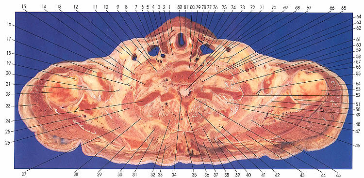
This section passes through the rima glottidis (82), cricoid cartilage (80), pharynx (81), first thoracic vertebra (72), elements of the brachial plexus (12, 67), and ribs 1 (16, 62) and 2 (24, 52).
The brachial plexus of nerves usually passes into the posterior triangle of the neck between the anterior (71) and middle (64) scalene muscles.
The inferior constrictor (2) is seen for the last time. The pharynx ends in this section; this occurs topographically at the level of the cricoid cartilage (80).
The subscapular artery, lateral thoracic branch, is seen supplying serratus anterior (55).
This variation occurs in about 30°o of individuals. The lateral thoracic artery has also been reported to arise from the second part of the axillary in 50%, from the first part of the axillary in 11%, and from the thoracoacromial artery in 7%.
The thyroid gland (4, 78) is seen bilaterally in this section. Muscles acting on the scapula in this section include serratus anterior (26, 47), rhomboideus minor (31, 39), rhomboideus major (30, 38), trapezius (33, 37), and deltoid (23, 54).
Muscles arising from the scapula in this section include subscapularis (27, 45) and infraspinatus (29, 43).
Next Page | Previous Page | Section Top | Title Page
Please send us comments by filling out our Comment Form.
Anatomy Atlases is licensed under a Creative Commons Attribution-NonCommercial-ShareAlike 4.0 International License. 
"Anatomy Atlases", the Anatomy Atlases logo, and "A digital library of anatomy information" are all Trademarks of Michael P. D'Alessandro, M.D.
Anatomy Atlases is funded in whole by Michael P. D'Alessandro, M.D. Advertising is not accepted.
Your personal information remains confidential and is not sold, leased, or given to any third party be they reliable or not.
The information contained in Anatomy Atlases is not a substitute for the medical care and advice of your physician. There may be variations in treatment that your physician may recommend based on individual facts and circumstances.
URL: http://www.anatomyatlases.org/