
Atlas of Human Anatomy in Cross Section: Section 2. Neck, Shoulders, Upper Arm, and Upper Thorax (Lungs)
Ronald A. Bergman, Ph.D., Adel K. Afifi, M.D., Jean J. Jew, M.D., and Paul
C. Reimann, B.S.
Peer Review Status: Externally Peer Reviewed
|
Upper Left Quadrant |
Lower Left Quadrant |
|
1. Anterior jugular v. |
30. Triceps brachii, lateral head m. |
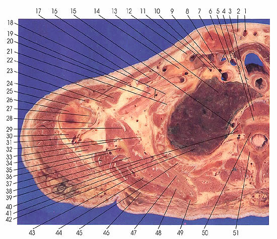
This section contains numerous important arterial and venous vessels. Arterial vessels include the inferior thyroid (110), common carotid (5, 107), internal thoracic (11, 103), subclavian (9, 101), circumflex scapular (33, G8), posterior circumflex humeral (31), and thoracoacromial (19). Identifiable veins include the internal jugular and subclavian (6, 8, 105,108), external jugular (8,104), suprascapular (36,102), axillary (14,88,100), transverse cervical (97), cephalic (91), circumflex scapular (33, 68), posterior circumflex humeral (31), thoracoacromial (19), azygos (15), and anterior jugular (1, 11).
The left lung bronchopulmonary segments are the left upper lobe and division, anterior segment (99), and left upper lobe and division, apical posterior segment (70). The right
lung segments include the right upper lobe, posterior segment (42), and right upper lobe, apical segment (13).
The brachial plexus of nerves is seen in the axilla (18, 86). The vagus (6, 107) and recurrent laryngeal (3, 110) nerves are seen. Note the position of the recurrent nerves between the esophagus (111) and trachea (112).
Muscles associated with the scapula include the serratus anterior (47,67), subscapularis (40, 62), teres major (28, 78), teres minor (37, 66), infraspinatus (43, 63), rhomboideus major (58), and the long head of the triceps muscles (30, 73).
The thyroid gland isthmus usually lies at the level of the seventh cervical vertebra in the true transverse plane. This section is slightly slanted so that the organ appears to lie at a level between the second and third thoracic vertebrae.
Next Page | Previous Page | Section Top | Title Page
Please send us comments by filling out our Comment Form.
Anatomy Atlases is licensed under a Creative Commons Attribution-NonCommercial-ShareAlike 4.0 International License. 
"Anatomy Atlases", the Anatomy Atlases logo, and "A digital library of anatomy information" are all Trademarks of Michael P. D'Alessandro, M.D.
Anatomy Atlases is funded in whole by Michael P. D'Alessandro, M.D. Advertising is not accepted.
Your personal information remains confidential and is not sold, leased, or given to any third party be they reliable or not.
The information contained in Anatomy Atlases is not a substitute for the medical care and advice of your physician. There may be variations in treatment that your physician may recommend based on individual facts and circumstances.
URL: http://www.anatomyatlases.org/