
Atlas of Human Anatomy in Cross Section: Section 2. Neck, Shoulders, Upper Arm, and Upper Thorax (Lungs)
Ronald A. Bergman, Ph.D., Adel K. Afifi, M.D., Jean J. Jew, M.D., and Paul
C. Reimann, B.S.
Peer Review Status: Externally Peer Reviewed
|
Upper Left Quadrant |
Lower Left Quadrant |
|
1. Vertebral plexus of vv., anterior |
32. Triceps brachii, lateral head m. |
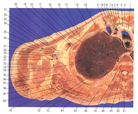
This section passes through the manubrium (5, 103), sternoclavicular joints (9, 10, 101), trachea (105), esophagus (104), mesoesophagus (93), several bronchopulmonary segments (17, 42, 66, 95), the third (2) and fourth (48) thoracic vertebrae, scapulae (37, 40, 45, 56, 61, 64), and the humeri (30, 79).
The lungs are the dominant structures at this level. The following bronchopulmonary segments are identifiable: left lung, upper lobe and division, anterior segment (95) and apical posterior segment (66), and right lung, upper lobe, posterior (42) and apical (17) segments.
The muscles of the arm include the pectoralis minor (16, 92), biceps brachii, long (85) and short (84) heads, pectoralis major (13, 20, 23, 83), coracobrachialis (25, 82), latissimus dorsi (69, 80), deltoid (70, 74, 78), triceps brachii, lateral (32, 72) and long (36, 71) heads, teres major (34), subscapularis (44, 58, 68), teres minor (38, 63), and infraspinatus (41).
The nerves of the brachial plexus (22, 27, 28, 73, 75 77) are seen in the axilla.
The common carotid (6) and right subclavian (8) arteries are seen at their bifurcation from the right brachiocephalic trunk.
Next Page | Previous Page | Section Top | Title Page
Please send us comments by filling out our Comment Form.
Anatomy Atlases is licensed under a Creative Commons Attribution-NonCommercial-ShareAlike 4.0 International License. 
"Anatomy Atlases", the Anatomy Atlases logo, and "A digital library of anatomy information" are all Trademarks of Michael P. D'Alessandro, M.D.
Anatomy Atlases is funded in whole by Michael P. D'Alessandro, M.D. Advertising is not accepted.
Your personal information remains confidential and is not sold, leased, or given to any third party be they reliable or not.
The information contained in Anatomy Atlases is not a substitute for the medical care and advice of your physician. There may be variations in treatment that your physician may recommend based on individual facts and circumstances.
URL: http://www.anatomyatlases.org/