
Atlas of Human Anatomy in Cross Section: Section 2. Neck, Shoulders, Upper Arm, and Upper Thorax (Lungs)
Ronald A. Bergman, Ph.D., Adel K. Afifi, M.D., Jean J. Jew, M.D., and Paul
C. Reimann, B.S.
Peer Review Status: Externally Peer Reviewed
|
Upper Left Quadrant |
Lower Left Quadrant |
|
1. Brachiocephalic v., right |
17. Radial nerve |
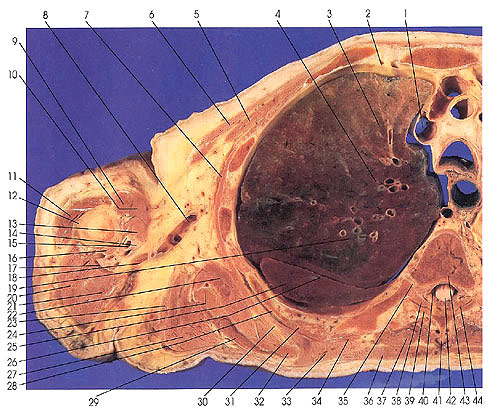
This section passes through the aorta (78) and its three primary arterial branches: right brachiocephalic trunk (86) and left common carotid (83) and left subclavian (80) arteries. The trachea (85) is seen next to a dilated or open esophagus (82). The section cuts the sixth thoracic vertebra (43).
The esophagus is the first segment of the alimentary canal and extends from the pharynx to the stomach. The esophagus may be divided into three segments: cervical, thoracic, and abdominal. The esophagus in the cervical and abdominal regions is generally closed, but the thoracic (as seen here) is more or less open, containing air.
Several bronchopulmonary segments can be recognized: left lung, upper lobe and division, anterior (79) and apical posterior (57) segments, and right lung, upper lobe, posterior (19), apical (4), and anterior (3) segments.
Next Page | Previous Page | Section Top | Title Page
Please send us comments by filling out our Comment Form.
Anatomy Atlases is licensed under a Creative Commons Attribution-NonCommercial-ShareAlike 4.0 International License. 
"Anatomy Atlases", the Anatomy Atlases logo, and "A digital library of anatomy information" are all Trademarks of Michael P. D'Alessandro, M.D.
Anatomy Atlases is funded in whole by Michael P. D'Alessandro, M.D. Advertising is not accepted.
Your personal information remains confidential and is not sold, leased, or given to any third party be they reliable or not.
The information contained in Anatomy Atlases is not a substitute for the medical care and advice of your physician. There may be variations in treatment that your physician may recommend based on individual facts and circumstances.
URL: http://www.anatomyatlases.org/