
Atlas of Human Anatomy in Cross Section: Section 4. Upper Limb
Ronald A. Bergman, Ph.D., Adel K. Afifi, M.D., Jean J. Jew, M.D., and Paul
C. Reimann, B.S.
Peer Review Status: Externally Peer Reviewed
|
Upper Left Quadrant |
Lower Left Quadrant |
Lower Right Quadrant |
Upper Right Quadrant |
|
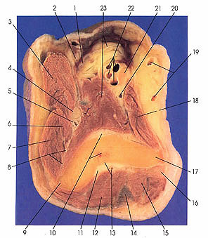
1. Tendon m. biceps brachii |
5. Radial collateral a. |
12. Triceps brachii m. |
18. Pronator teres m. (first appearance) |
This section passes through the lateral (9) and medial (17) epicondyles of the humerus. Note that the ulnar nerve (16) lies in the ulnar sulcus on the posterior side of the medial epicondyle (17). The radial nerve (4) is located between the brachioradialis (3), brachialis (23), and extensor carpi radialis brevis (6) muscles. The radial nerve supplies all three muscles. The primary innervation of brachialis, however, is the musculocutaneous nerve represented at this level by its cutaneous branch, the lateral antebrachial cutaneous nerve (2). The median nerve (21) is steadfast in its position adjacent to the brachial artery and vein (22).
The radial collateral (5) and the middle collateral (13) arteries are seen in this cut. They are branches of the deep brachial artery. These vessels anastomose with the radial recurrent and the interosseous recurrent arteries, respectively. The inferior ulnar collateral artery (20), also a part of the anastomotic network of blood vessels around the elbow, is also seen.
An extensive hematoma is seen in the anterior superficial (fatty) fascia.
Note the origin of extensor carpi radialis brevis (6) (first appearance) and extensor carpi radialis longus (7) from the lateral intermuscular septum (8), which extends from the lower third of the lateral epicondylar ridge (9). Pronator teres (18) is seen for the first time.
Next Page | Previous Page | Section Top | Title Page
Please send us comments by filling out our Comment Form.
Anatomy Atlases is licensed under a Creative Commons Attribution-NonCommercial-ShareAlike 4.0 International License. 
"Anatomy Atlases", the Anatomy Atlases logo, and "A digital library of anatomy information" are all Trademarks of Michael P. D'Alessandro, M.D.
Anatomy Atlases is funded in whole by Michael P. D'Alessandro, M.D. Advertising is not accepted.
Your personal information remains confidential and is not sold, leased, or given to any third party be they reliable or not.
The information contained in Anatomy Atlases is not a substitute for the medical care and advice of your physician. There may be variations in treatment that your physician may recommend based on individual facts and circumstances.
URL: http://www.anatomyatlases.org/