
Atlas of Human Anatomy in Cross Section: Section 4. Upper Limb
Ronald A. Bergman, Ph.D., Adel K. Afifi, M.D., Jean J. Jew, M.D., and Paul
C. Reimann, B.S.
Peer Review Status: Externally Peer Reviewed
|
Upper Left Quadrant |
Lower Left Quadrant |
Lower Right Quadrant |
Upper Right Quadrant |
|
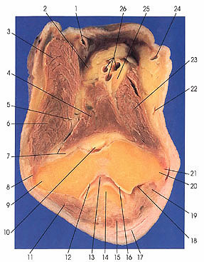
1. Lateral antebrachial cutaneous nerve and cephalic v. |
7. Radial nerve, deep br. and radial fossa |
15. Tendon m. triceps brachii |
22. Anastomotic v. |
This section passes through the medial (20) and lateral (9) epicondyles of the humerus, the trochlea (12, 16), the ulnar nerve sulcus (18), the coronoid (10) and radial (7) fossae of the humerus, and the tip of the olecranon process of the ulna (14). It cuts the anconeus muscle (11), which appears for the first time.
The median nerve (25) is moving into the space between pronator teres (23) and brachialis (4) muscles and will enter the forearm beneath a fibrous arch that separates the humeral and ulnar or deep heads of pronator teres. It will lie between the flexor digitorum superficialis and flexor digitorum profundus muscles before reaching the hand. In its course, it supplies all flexor muscles except the medial half of flexor digitorum profundus and none of flexor carpi ulnaris. These are supplied by the ulnar nerve (19).
The radial nerve has divided into its two terminal branches; the deep branch (7) supplies extensor carpi radialis brevis, supinator, extensor digitorum communis, extensor digiti minimi, extensor carpi ulnaris, extensor indicts, extensor pollicis longus, extensor pollicis brevis, and abductor pollicis longus muscles. The superficial branch (5) is purely sensory to the back of the forearm, wrist, and the hand. Its most lateral branch covers the area of the radial side of the thenar eminence. On its medial side, it intermingles with the ulnar nerve. Other branches are called dorsal digital nerves. They supply a variable amount of skin on the dorsum of the first digit, both sides of the second digit, and the radial side of the third digit. These branches usually extend to the base of the nail of the first digit, to the distal interphalangeal joint of the second, just short of the proximal interphalangeal joint of the third, and to the metacarpophalangeal joint of the fourth digit.
Next Page | Previous Page | Section Top | Title Page
Please send us comments by filling out our Comment Form.
Anatomy Atlases is licensed under a Creative Commons Attribution-NonCommercial-ShareAlike 4.0 International License. 
"Anatomy Atlases", the Anatomy Atlases logo, and "A digital library of anatomy information" are all Trademarks of Michael P. D'Alessandro, M.D.
Anatomy Atlases is funded in whole by Michael P. D'Alessandro, M.D. Advertising is not accepted.
Your personal information remains confidential and is not sold, leased, or given to any third party be they reliable or not.
The information contained in Anatomy Atlases is not a substitute for the medical care and advice of your physician. There may be variations in treatment that your physician may recommend based on individual facts and circumstances.
URL: http://www.anatomyatlases.org/