
Atlas of Human Anatomy in Cross Section: Section 4. Upper Limb
Ronald A. Bergman, Ph.D., Adel K. Afifi, M.D., Jean J. Jew, M.D., and Paul
C. Reimann, B.S.
Peer Review Status: Externally Peer Reviewed
|
Upper Left Quadrant |
Lower Left Quadrant |
Lower Right Quadrant |
Upper Right Quadrant |
|
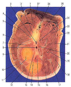
1. Radial nerve, superficial br. |
8. Ulnar a. |
15. Brachialis m. |
21. Palmaris longus m. |
This section passes through the radial neck (11) and the ulnar tuberosity (14). The median nerve (24) supplies all of the muscles of the flexor forearm except for flexor carpi ulnaris (18) and the ulnar half of flexor digitorum profundus (16). These are supplied by the ulnar nerve (17).
The superficial branch of the radial nerve (1) is purely sensory and may be found running under cover of brachioradialis (27) anterior to the elbow joint. It penetrates the forearm and appears on the back of the forearm where its most lateral branches supply the radial part of the thenar eminence. The most medial branch mingles with terminal branches of the ulnar nerve and is called the ulnar anastomotic branch. The deep branch of the radial nerve (4) becomes known as the posterior interosseous nerve when it enters the space between the superficial and deep muscles of the back of the forearm.
The deep branch of the radial nerve supplies the following muscles: extensor carpi radialis brevis (6), supinator (7), extensor digitorum communis (9), extensor digiti minimi, extensor carpi ulnaris ( 10), extensor indicis, extensor pollicis longus, extensor pollicis brevis, and abductor pollicis longus.
The deep branch, after becoming the posterior interosseous nerve, accompanies the posterior interosseous artery running along the posterior interosseous membrane and back of the radius. At the wrist the nerve passes in the groove for extensor digitorum communis and extensor indicis muscles and terminates as sensory branches to the carpal joints.
Brachialis (15) makes its last appearance in this section.
Next Page | Previous Page | Section Top | Title Page
Please send us comments by filling out our Comment Form.
Anatomy Atlases is licensed under a Creative Commons Attribution-NonCommercial-ShareAlike 4.0 International License. 
"Anatomy Atlases", the Anatomy Atlases logo, and "A digital library of anatomy information" are all Trademarks of Michael P. D'Alessandro, M.D.
Anatomy Atlases is funded in whole by Michael P. D'Alessandro, M.D. Advertising is not accepted.
Your personal information remains confidential and is not sold, leased, or given to any third party be they reliable or not.
The information contained in Anatomy Atlases is not a substitute for the medical care and advice of your physician. There may be variations in treatment that your physician may recommend based on individual facts and circumstances.
URL: http://www.anatomyatlases.org/