
Atlas of Human Anatomy in Cross Section: Section 4. Upper Limb
Ronald A. Bergman, Ph.D., Adel K. Afifi, M.D., Jean J. Jew, M.D., and Paul
C. Reimann, B.S.
Peer Review Status: Externally Peer Reviewed
|
Upper Left Quadrant |
Lower Left Quadrant |
Lower Right Quadrant |
Upper Right Quadrant |
|
1. Tendon m. palmaris longus |
7. Brachioradialis m. |
14. Abductor pollicis longus m. |
21. Basilic v. |
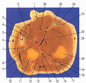
This section passes through the middle of the forearm.
The radiodorsal or extensor muscles lie in two layers, superficial and deep. The muscles of the superficial layer on the radial side, or radial set, consist of three muscles: brachioradialis (7), extensor carpi radialis longus (9), and extensor carpi radialis brevis (10). Brachioradialis arises from the lateral epicondylar ridge of the humerus and inserts onto the styloid process of the radius. Extensor carpi radialis longus arises from the lateral epicondylar ridge of the humerus and inserts onto the second metacarpal bone. Extensor carpi radialis brevis arises from the lateral epicondylar ridge and inserts onto the third metacarpal. These muscles are innervated by branches of the radial nerve that arise proximal to the passage of the deep radial (posterior interosseous) through the supinator muscle. The intermediate set of muscles of the superficial layer consists of extensor digitorum (13) and extensor digiti minimi (16) muscles. They arise from the lateral epicondyle of the humerus and are inserted onto the backs of the fingers. The ulnar set consists of extensor carpi ulnaris (19), which arises from the lateral epicondyle of the humerus and is inserted onto the ulnar side of the base of the fifth metacarpal. The intermediate and ulnar sets of muscles are supplied by branches from the deep ramus of the radial nerve after it has passed through the supinator muscle.
The deep layer is composed of supinator, abductor pollicis longus ( 14), extensor pollicis brevis, and extensor indicis muscles. The latter two muscles are seen in subsequent sections. Supinator was seen previously. This rhomboid shaped muscle arises from the lateral epicondyle of the humerus and supinator crest of the ulna, winds laterally around the radius, and inserts on its palmer surface. Abductor pollicis longus (14), a fusiform muscle, arises from the middle third of the ulna, interosseous membrane, and radius and inserts onto the base of the first metacarpal. Extensor pollicis brevis arises from the radius distal to abductor pollicis longus and inserts onto the base of rhe proximal phalanx of the thumb. Extensor pollicis longus is a narrow muscle that arises from the middle third of the dorsal surface of the ulna and inserts onto the base of the distal phalanx of the thumb. Extensor indicis is a narrow fusiform muscle arising from the shaft of the ulna and inserts onto the dorsal aponeurosis of the index finger. These muscles are innervated by branches of the deep radial (posterior interosseous) nerve while it is passing through or after it passes through the supinator muscle.
Next Page | Previous Page | Section Top | Title Page
Please send us comments by filling out our Comment Form.
Anatomy Atlases is licensed under a Creative Commons Attribution-NonCommercial-ShareAlike 4.0 International License. 
"Anatomy Atlases", the Anatomy Atlases logo, and "A digital library of anatomy information" are all Trademarks of Michael P. D'Alessandro, M.D.
Anatomy Atlases is funded in whole by Michael P. D'Alessandro, M.D. Advertising is not accepted.
Your personal information remains confidential and is not sold, leased, or given to any third party be they reliable or not.
The information contained in Anatomy Atlases is not a substitute for the medical care and advice of your physician. There may be variations in treatment that your physician may recommend based on individual facts and circumstances.
URL: http://www.anatomyatlases.org/