
Atlas of Human Anatomy in Cross Section: Section 4. Upper Limb
Ronald A. Bergman, Ph.D., Adel K. Afifi, M.D., Jean J. Jew, M.D., and Paul
C. Reimann, B.S.
Peer Review Status: Externally Peer Reviewed
|
Upper Left Quadrant |
Lower Left Quadrant |
Lower Right Quadrant |
Upper Right Quadrant |
|
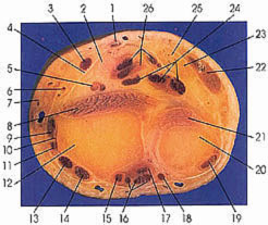
1. Tendon m. palmaris longus |
9. Tendon m. abductor pollicis longus |
17. Extensor indicis m. and tendon |
22. Flexor carpi ulnaris m. and tendon |
In this section pronator quadratus (8, 21) is thinning and is seen for the last time. All other muscles are tendinous.
The radius is still expanding and will ultimately articulate with three carpal bones. The head of the ulna (20) is beginning to articulate with the radius (12). Note the articular cartilage at its medial border (articular surface) adjacent to the radius.
The tendons of flexor digitorum superficialis (26) and profundus (24) and flexor pollicis
longus (5) and the median nerve (2) are already enclosed within a fascial covering before entering the carpal tunnel. Flexor carpi radialis (3) enters the hand through its own osteofibrous tunnel formed by the attachment of the flexor retinaculum to the two borders of the groove on the trapezium. The tendon of flexor carpi radialis (3) is already separated by a fascial sheath (4) from the tendons that enter the carpal tunnel.
Next Page | Previous Page | Section Top | Title Page
Please send us comments by filling out our Comment Form.
Anatomy Atlases is licensed under a Creative Commons Attribution-NonCommercial-ShareAlike 4.0 International License. 
"Anatomy Atlases", the Anatomy Atlases logo, and "A digital library of anatomy information" are all Trademarks of Michael P. D'Alessandro, M.D.
Anatomy Atlases is funded in whole by Michael P. D'Alessandro, M.D. Advertising is not accepted.
Your personal information remains confidential and is not sold, leased, or given to any third party be they reliable or not.
The information contained in Anatomy Atlases is not a substitute for the medical care and advice of your physician. There may be variations in treatment that your physician may recommend based on individual facts and circumstances.
URL: http://www.anatomyatlases.org/