
Atlas of Human Anatomy in Cross Section: Section 4. Upper Limb
Ronald A. Bergman, Ph.D., Adel K. Afifi, M.D., Jean J. Jew, M.D., and Paul
C. Reimann, B.S.
Peer Review Status: Externally Peer Reviewed
|
Upper Left Quadrant |
Lower Left Quadrant |
Lower Right Quadrant |
Upper Right Quadrant |
|
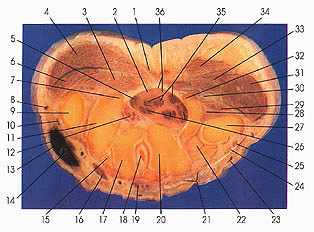
1. Tendon m. palmaris longus |
9. First metacarpal base |
20. Carpal bone, capitate |
29. Flexor digiti minimi brevis m. |
This is the last section through the carpal canal and the dense flexor retinaculum (36). The retinaculum passes between the hook of the hamate (32) and the crest or tubercle of the trapezium (7).
The ulnar nerve (30) is seen passing medial to the hook of the hamate (32) to enter the hand. The deep branch of the ulnar nerve usually, but not always, supplies the three hypothenar muscles (31, 33), all seven interossei, the third and fourth lumbricals, the adductor pollicis muscle, and several joints. The superficial branch supplies the palmaris
brevis muscle (34) and is sensory for the little finger and the medial side of the ring finger and the medial side of the palm of the hand.
The passage of the ulnar nerve, deep branch (30), into the hand is via a canal (of Guyon) whose sides (with the hand in the anatomic position) include the pisohamate ligament above, the pisiform bone medially, the hook of the hamate laterally, and the transverse carpal ligament below.
Next Page | Previous Page | Section Top | Title Page
Please send us comments by filling out our Comment Form.
Anatomy Atlases is licensed under a Creative Commons Attribution-NonCommercial-ShareAlike 4.0 International License. 
"Anatomy Atlases", the Anatomy Atlases logo, and "A digital library of anatomy information" are all Trademarks of Michael P. D'Alessandro, M.D.
Anatomy Atlases is funded in whole by Michael P. D'Alessandro, M.D. Advertising is not accepted.
Your personal information remains confidential and is not sold, leased, or given to any third party be they reliable or not.
The information contained in Anatomy Atlases is not a substitute for the medical care and advice of your physician. There may be variations in treatment that your physician may recommend based on individual facts and circumstances.
URL: http://www.anatomyatlases.org/