
Atlas of Human Anatomy in Cross Section: Section 4. Upper Limb
Ronald A. Bergman, Ph.D., Adel K. Afifi, M.D., Jean J. Jew, M.D., and Paul
C. Reimann, B.S.
Peer Review Status: Externally Peer Reviewed
|
Upper Left Quadrant |
Lower Left Quadrant |
Lower Right Quadrant |
Upper Right Quadrant |
|
1. Digital aa. and palmar digital nerve |
8. Adductor pollicis, transverse head m. |
16. Tendon m. extensor digitorum communis |
27. Tendon m. extensor digiti minimi |
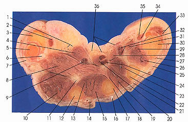
In this section, the four dorsal (10, 15, 19, 26) and three palmer (13, 23, 29) interosseous muscles are seen. When four palmer interosseous muscles are described the first palmer interosseous muscle is a division or part of flexor pollicis brevis or adductor pollicis. One origin of adductor pollicis (18) from the third metacarpal bone (17) can be seen. These muscles are all usually innervated by the deep palmer branch of the ulnar nerve. It is of some interest that flexor pollicis brevis is also supplied by the recurrent branch of the first palmer digital ramus of the median nerve as well as by the ulnar nerve. In one study, both nerves supplied the muscle in 19 of 29 cases, in five cases by the median alone, and in five cases by the ulnar alone.
All four lumbrical muscles are identified (4, 20, 28, 31). The first and second (4, 20) are usually median nerve innervated and the third and fourth (28, 31) are ulnar nerve innervated.
A sesamoid bone (6) of the thumb is seen.
Next Page | Previous Page | Section Top | Title Page
Please send us comments by filling out our Comment Form.
Anatomy Atlases is licensed under a Creative Commons Attribution-NonCommercial-ShareAlike 4.0 International License. 
"Anatomy Atlases", the Anatomy Atlases logo, and "A digital library of anatomy information" are all Trademarks of Michael P. D'Alessandro, M.D.
Anatomy Atlases is funded in whole by Michael P. D'Alessandro, M.D. Advertising is not accepted.
Your personal information remains confidential and is not sold, leased, or given to any third party be they reliable or not.
The information contained in Anatomy Atlases is not a substitute for the medical care and advice of your physician. There may be variations in treatment that your physician may recommend based on individual facts and circumstances.
URL: http://www.anatomyatlases.org/