
Atlas of Human Anatomy in Cross Section: Section 4. Upper Limb
Ronald A. Bergman, Ph.D., Adel K. Afifi, M.D., Jean J. Jew, M.D., and Paul
C. Reimann, B.S.
Peer Review Status: Externally Peer Reviewed
|
Upper Left Quadrant |
Lower Left Quadrant |
Lower Right Quadrant |
Upper Right Quadrant |
|
1. First dorsal metacarpal a. |
7. Tendon m. flexor digitorum superficialis |
17. Tendon m. extensor digitorum communis |
29. Fourth lumbrical m. |
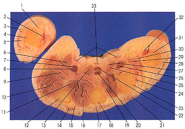
This section is beyond the junction of the thumb with the hand. The thumb is cut through the first or proximal phalanx (4).
The fourth lumbrical (30) is doubled, but this is considered a normal variation. The third and fourth lumbricals frequently show anatomic variation.
The four lumbricals arise from the tendons of flexor digitorum profundus and insert into the extensor expansion on the radial side of the corresponding finger. In one study of 110 bodies (220 hands), it was reported that all four lumbricals were inserted on the radial side of the extensor tendon on the dorsum of their respective digits in 39% of hands. In 35%, the first, second, and fourth were inserted on the radial side, while the third was inserted on adjacent sides of both the middle and ring fingers. The remaining 25% of hands exhibited other types of anomalous origins and insertions. An accessory fascicle was found to arise from flexor pollicis longus to insert on the index finger.
Next Page | Previous Page | Section Top | Title Page
Please send us comments by filling out our Comment Form.
Anatomy Atlases is licensed under a Creative Commons Attribution-NonCommercial-ShareAlike 4.0 International License. 
"Anatomy Atlases", the Anatomy Atlases logo, and "A digital library of anatomy information" are all Trademarks of Michael P. D'Alessandro, M.D.
Anatomy Atlases is funded in whole by Michael P. D'Alessandro, M.D. Advertising is not accepted.
Your personal information remains confidential and is not sold, leased, or given to any third party be they reliable or not.
The information contained in Anatomy Atlases is not a substitute for the medical care and advice of your physician. There may be variations in treatment that your physician may recommend based on individual facts and circumstances.
URL: http://www.anatomyatlases.org/