
Atlas of Human Anatomy in Cross Section: Section 6. Pelvis, Perineum, Hip, and Upper Thigh
Ronald A. Bergman, Ph.D., Adel K. Afifi, M.D., Jean J. Jew, M.D., and Paul
C. Reimann, B.S.
Peer Review Status: Externally Peer Reviewed
|
Upper Left Quadrant |
Lower Left Quadrant |
Lower Right Quadrant |
Upper Right Quadrant |
|
1. Median and lateral umbilical ligaments |
11. Common iliac v. |
25. Spinous process, L5 |
41. Genitofemoral nerve |
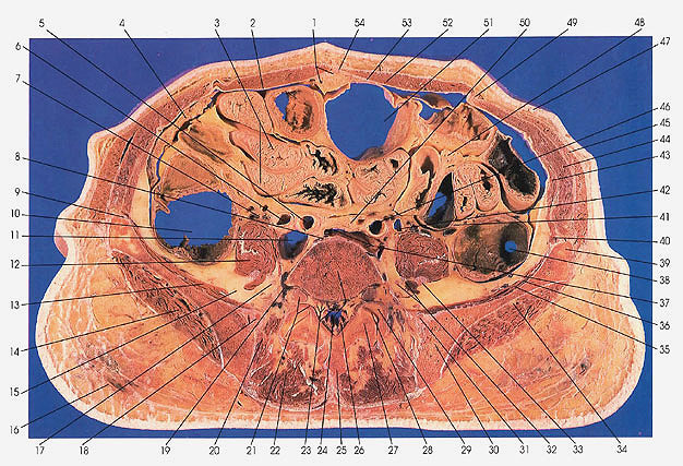
This section is through the middle of the fifth lumbar vertebra (24) at the inferior edge of the transverse process. The superior (29) and inferior (27) articular processes and the intervertebral joint (28) between L5 and S1 are seen.
The distal end of the inferior vena cave is seen branching to the left to form the left common iliac vein (37). The inferior vena cave (37) makes its last appearance at this level.
The right common iliac vein (11) is already present at this level.
The ureters (6, 42) can be seen flanking the common iliac arteries (5, 46).
The iliohypogastric nerve (9) is seen at the iliac crest (39), where it will leave the greater or abdominal pelvis and move anteriorly between the transverse abdominis and internal oblique muscles to the anterior abdominal wall.
Next Page | Previous Page | Section Top | Title Page
Please send us comments by filling out our Comment Form.
Anatomy Atlases is licensed under a Creative Commons Attribution-NonCommercial-ShareAlike 4.0 International License. 
"Anatomy Atlases", the Anatomy Atlases logo, and "A digital library of anatomy information" are all Trademarks of Michael P. D'Alessandro, M.D.
Anatomy Atlases is funded in whole by Michael P. D'Alessandro, M.D. Advertising is not accepted.
Your personal information remains confidential and is not sold, leased, or given to any third party be they reliable or not.
The information contained in Anatomy Atlases is not a substitute for the medical care and advice of your physician. There may be variations in treatment that your physician may recommend based on individual facts and circumstances.
URL: http://www.anatomyatlases.org/