
Atlas of Human Anatomy in Cross Section: Section 6. Pelvis, Perineum, Hip, and Upper Thigh
Ronald A. Bergman, Ph.D., Adel K. Afifi, M.D., Jean J. Jew, M.D., and Paul
C. Reimann, B.S.
Peer Review Status: Externally Peer Reviewed
|
Upper Left Quadrant |
Lower Left Quadrant |
Lower Right Quadrant |
Upper Right Quadrant |
|
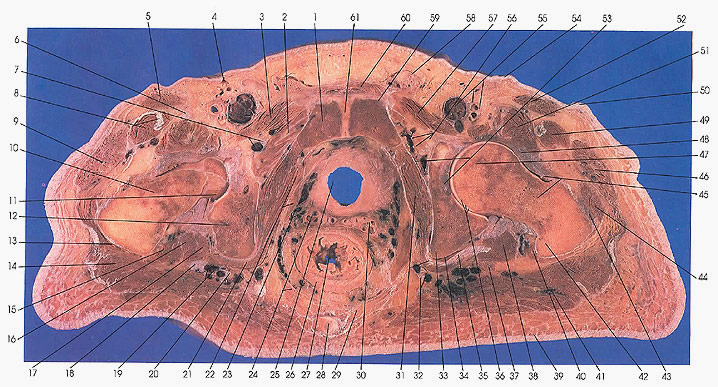
1. Pubis |
9. Tensor fascia lata m. |
28. Coccyx |
46. Tensor fascia lata m. |
This section passes through the coccyx (28), the lesser sciatic notch, the acetabulum (36), the head (47) and neck (45) of the femur on the left side and the inferior edge of the acetabulum on the right side. The cut passes through the obturator foramen (56) and membrane (53) and the pubic symphysis (61).
Note the relationship between the inferior gluteal blood vessels (19), sciatic nerve (18, 37), the tendon of the obturator internus muscle (21, 35), the gemelli muscles (15, 17, 38), quadratus femoris (40), and the gluteus maximus muscle (14, 41). Recall that all these structures receive their blood supply, in part or entirely, from branches of the inferior gluteal vessels.
The inferior gluteal artery also assists in forming the cruciate anastomosis that forms around the neck (10, 45) and greater trochanter (42) of the femur. This involves the first perforating and lateral and medial femoral circumflex branches of the femoral artery (55) in addition to the inferior gluteal artery (19). The inferior gluteal vessels arise from the internal iliac vessels and traverse the lesser sciatic foremen (33) to leave and enter the pelvis.
The quadratus femoris (40) and obturator externus (2) muscles appear for the first time in this section.
Note the relationship between the obturator nerve (7, 56) and blood vessels (7, 56) and the obturator externus (2) and pectineus (3, 57) muscles. The obturator nerve innervates and obturator blood vessels supply, among others, these two muscles.
The trochanteric bursa of the gluteus maximus muscle can be seen.
Compare the thickness of the skin on the ventral surface (54) of the body with that on the dorsal surface (39).
Next Page | Previous Page | Section Top | Title Page
Please send us comments by filling out our Comment Form.
Anatomy Atlases is licensed under a Creative Commons Attribution-NonCommercial-ShareAlike 4.0 International License. 
"Anatomy Atlases", the Anatomy Atlases logo, and "A digital library of anatomy information" are all Trademarks of Michael P. D'Alessandro, M.D.
Anatomy Atlases is funded in whole by Michael P. D'Alessandro, M.D. Advertising is not accepted.
Your personal information remains confidential and is not sold, leased, or given to any third party be they reliable or not.
The information contained in Anatomy Atlases is not a substitute for the medical care and advice of your physician. There may be variations in treatment that your physician may recommend based on individual facts and circumstances.
URL: http://www.anatomyatlases.org/