
Atlas of Human Anatomy in Cross Section: Section 6. Pelvis, Perineum, Hip, and Upper Thigh
Ronald A. Bergman, Ph.D., Adel K. Afifi, M.D., Jean J. Jew, M.D., and Paul
C. Reimann, B.S.
Peer Review Status: Externally Peer Reviewed
|
Upper Left Quadrant |
Lower Left Quadrant |
Lower Right Quadrant |
Upper Right Quadrant |
|
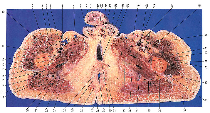
1. Penile septum (between corpora cavernosa) |
13. Adductor brevis m. |
27. Anal crease (crena ani) |
40. Tensor fascia lata m. |
This section passes through the penis (1), spermatic cords (2, 52), the corpus spongiosum (55) and bulbocavernosus muscle (51), the superficial transverse perineal muscle (30), the anal crease (27), and the anococcygeal ligament (28).
The pudendal plexus of veins (53) and pampiniform plexus (2, 52) are seen. The internal (24) and external (25) anal sphincters are identified.
The ischium (32) makes its last appearance in this section.
The femoral nerve is now seen divided into several branches (10, 46), which will supply, among others, the rectus femoris (9), sartorius (7), vastus intermedius (43), vastus medialis (42), and vastus lateralis (41).
Next Page | Previous Page | Section Top | Title Page
Please send us comments by filling out our Comment Form.
Anatomy Atlases is licensed under a Creative Commons Attribution-NonCommercial-ShareAlike 4.0 International License. 
"Anatomy Atlases", the Anatomy Atlases logo, and "A digital library of anatomy information" are all Trademarks of Michael P. D'Alessandro, M.D.
Anatomy Atlases is funded in whole by Michael P. D'Alessandro, M.D. Advertising is not accepted.
Your personal information remains confidential and is not sold, leased, or given to any third party be they reliable or not.
The information contained in Anatomy Atlases is not a substitute for the medical care and advice of your physician. There may be variations in treatment that your physician may recommend based on individual facts and circumstances.
URL: http://www.anatomyatlases.org/