
Atlas of Human Anatomy in Cross Section: Section 7. Lower Limb
Ronald A. Bergman, Ph.D., Adel K. Afifi, M.D., Jean J. Jew, M.D., and Paul
C. Reimann, B.S.
Peer Review Status: Externally Peer Reviewed
|
Upper Left Quadrant |
Lower Left Quadrant |
Lower Right Quadrant |
Upper Right Quadrant |
|
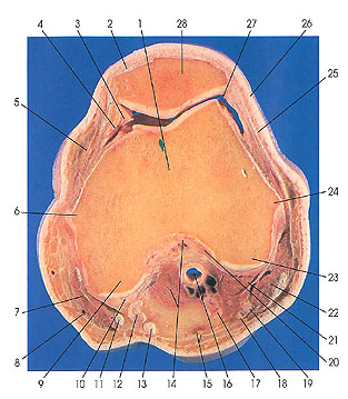
1. Femur |
6. Medial epicondyle |
14. Gastrocnemius m. and intercondylar fossa |
24. Lateral femoral epicondyle |
This section passes through the femoral condyles (6, 23) and the center of the patella (28).
Several important ligamentous structures are shown, including the iliotibial tract (25), anterior cruciate ligament (20) (for the first time), and the transverse medial patellar retinaculum (4). The large number of muscle tendons, including those of vastus lateralis (26), semitendinosus (13), semimembranosus (12), gracilis (11), vastus medialis (5), and the quadriceps femoris (2), are indicators of the proximity of the knee joint region.
The patella (28) is a large sesamoid bone related to those found in the hand and foot, which more closely in size resemble the sesame grain for which they are named. The patella consists of dense, spongy bone covered by a thin, compact layer of bone. The patella, by virtue of its position, performs the very important function of protecting the knee joint from injury. The patella's primary nutrient vessels are branches from the articular branch of the descending artery of the knee (descending genicular), the anterior tibial recurrent, and the articular rete of the knee.
The anterior cruciate ligament (20), seen for the first time in this cut, is very strong and cordlike. It is attached in the anterior intercondylar area of the tibia and to the lateral margin of the medial articular surface. It extends to the posterior part of the medial surface of the lateral condyle of the femur. It is fixed to the tibia posterior to the anterior extremity of the medial meniscus. Posteriorly and laterally is the anterior extremity of the lateral meniscus, from which some fibers blend with the lateral edge of the anterior cruciate ligament.
Next Page | Previous Page | Section Top | Title Page
Please send us comments by filling out our Comment Form.
Anatomy Atlases is licensed under a Creative Commons Attribution-NonCommercial-ShareAlike 4.0 International License. 
"Anatomy Atlases", the Anatomy Atlases logo, and "A digital library of anatomy information" are all Trademarks of Michael P. D'Alessandro, M.D.
Anatomy Atlases is funded in whole by Michael P. D'Alessandro, M.D. Advertising is not accepted.
Your personal information remains confidential and is not sold, leased, or given to any third party be they reliable or not.
The information contained in Anatomy Atlases is not a substitute for the medical care and advice of your physician. There may be variations in treatment that your physician may recommend based on individual facts and circumstances.
URL: http://www.anatomyatlases.org/