
Atlas of Human Anatomy in Cross Section: Section 7. Lower Limb
Ronald A. Bergman, Ph.D., Adel K. Afifi, M.D., Jean J. Jew, M.D., and Paul
C. Reimann, B.S.
Peer Review Status: Externally Peer Reviewed
|
Upper Left Quadrant |
Lower Left Quadrant |
Lower Right Quadrant |
Upper Right Quadrant |
|
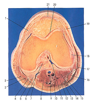
1. Medial patellar retinaculum |
2. Great saphenous v. |
9. Popliteal v. and a. |
19. Lateral patellar retinaculum |
This section passes through the patellar ligament (21) for the first time. Important ligaments also identified for the first time include the posterior cruciate (6) and the medial patellar retinaculum (1). Note that the sartorius muscle is becoming tendinous and that this begins on its medial surface, which is adjacent to the medial femoral condyle.
The posterior cruciate ligament (6) is stronger and less oblique than the anterior (11) one. It is fixed distally to the greater portion of the posterior intercondylar area, especially the lateral and posterior portions, being joined by fibers that are attached between the intercondylar tubercles. It extends to the anterior part of the lateral surface of the medial condyle of the femur, having a wide crescent-like attachment, about 1.5 cm in width, just proximal to the anterior surface. At the tibia, posteriorly, it is joined directly with the oblique ligament; anteriorly, it passes proximal to the posterior extremity of the medial meniscus; laterally, it receives slips from the lateral meniscus and from the anterior and posterior meniscofemoral ligaments, which join it anteriorly and posteriorly and extend with it to be attached to the femur. At the level of the intercondylar eminence of the tibia, the two cruciate ligaments are bound together so that no space exists between their tibial attachments and the point of their decussation.
The medial patellar retinaculum (1) is derived from the tendon of vastus medialis muscle. It is attached to the patella along its medial border, as far as the attachment of the patellar ligament, and passing along its sides to the tibia it attaches to an oblique ridge and extends as far as the tibial collateral ligament. Fibers of the medial patellar retinaculum extend distal to the oblique ridge to blend with the periosteum of the shaft. Both patellar retinacula become inseparably joined with the fibrous membrane of the articular capsule.
Next Page | Previous Page | Section Top | Title Page
Please send us comments by filling out our Comment Form.
Anatomy Atlases is licensed under a Creative Commons Attribution-NonCommercial-ShareAlike 4.0 International License. 
"Anatomy Atlases", the Anatomy Atlases logo, and "A digital library of anatomy information" are all Trademarks of Michael P. D'Alessandro, M.D.
Anatomy Atlases is funded in whole by Michael P. D'Alessandro, M.D. Advertising is not accepted.
Your personal information remains confidential and is not sold, leased, or given to any third party be they reliable or not.
The information contained in Anatomy Atlases is not a substitute for the medical care and advice of your physician. There may be variations in treatment that your physician may recommend based on individual facts and circumstances.
URL: http://www.anatomyatlases.org/