
Atlas of Human Anatomy in Cross Section: Section 7. Lower Limb
Ronald A. Bergman, Ph.D., Adel K. Afifi, M.D., Jean J. Jew, M.D., and Paul
C. Reimann, B.S.
Peer Review Status: Externally Peer Reviewed
|
Upper Left Quadrant |
Lower Left Quadrant |
Lower Right Quadrant |
Upper Right Quadrant |
|
1. Anterior cruciate ligament |
3. Lateral femoral condyle, articular cartilage |
11. Popliteal v. and a. |
22. Tibial collateral ligament |
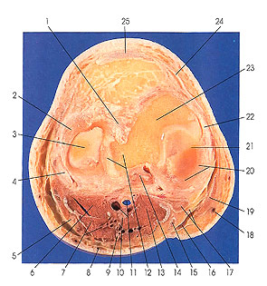
This section is the superior face of the next section (Plate 7.15); you are looking downward (distally) rather than upward.
The section passes through the knee joint patellar ligament (2 5), tibial (22) and fibular (4) collateral ligaments, the medial (20) and lateral (2) menisci, the anterior (1) and posterior (14) cruciate ligaments, and the distal edges of the medial (21) and lateral (3) femoral condyles.
It cuts the tibial nerve (10), medial sural cutaneous nerve (9), nerve to the soleus (10), nerve to gastrocnemius (lateral head) (7), and the common peroneal nerve (5).
Plantaris muscle (14) makes its first appearance in this section.
The tibial collateral ligament (22) is a strong, flat band that extends from the tubercle on the medial epicondyle of the femur to the medial margin and medial surface of the shaft of the tibia, about 3.5 cm distal to the condyle. The fibular collateral ligament (4) is a strong round cord, about 5 cm in length, attached proximally to a tubercle on the lateral epicondyle of the femur and fixed distally to the middle of the lateral surface of the head of the fibula about 1.5 cm from the apex.
The menisci (2, 20) are two intraarticular crescentic disks of fibrocartilage resting on circular portions of the articular surface of the tibia. They move with the tibia upon the femur. They deepen the shallow tibial articular surfaces. They are dense and compact in structure, becoming looser and more fibrous near their extremities, where they are fixed firmly both anteriorly and posteriorly to the intercondylar eminence of the tibia. The circumferential border of each disk is convex, thick, and rather loosely attached to the margins of the condyles of the tibia by the fibrous membrane of the capsule. The inner margins are concave, thin, and free. They are about 1.3 to 1.5 cm broad at their widest part; they taper toward their extremities and cover less than two-thirds of the articular surface of the tibia. Their proximal surfaces are slightly concave; the distal surfaces are flat and rest upon the tibia.
Next Page | Previous Page | Section Top | Title Page
Please send us comments by filling out our Comment Form.
Anatomy Atlases is licensed under a Creative Commons Attribution-NonCommercial-ShareAlike 4.0 International License. 
"Anatomy Atlases", the Anatomy Atlases logo, and "A digital library of anatomy information" are all Trademarks of Michael P. D'Alessandro, M.D.
Anatomy Atlases is funded in whole by Michael P. D'Alessandro, M.D. Advertising is not accepted.
Your personal information remains confidential and is not sold, leased, or given to any third party be they reliable or not.
The information contained in Anatomy Atlases is not a substitute for the medical care and advice of your physician. There may be variations in treatment that your physician may recommend based on individual facts and circumstances.
URL: http://www.anatomyatlases.org/