
Atlas of Human Anatomy in Cross Section: Section 7. Lower Limb
Ronald A. Bergman, Ph.D., Adel K. Afifi, M.D., Jean J. Jew, M.D., and Paul
C. Reimann, B.S.
Peer Review Status: Externally Peer Reviewed
|
Upper Left Quadrant |
Lower Left Quadrant |
Lower Right Quadrant |
Upper Right Quadrant |
|
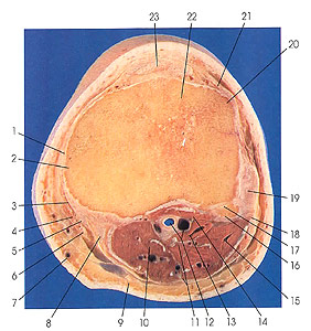
1. Medial patellar retinaculum |
3. Anserine bursa |
11. Medial sural cutaneous and tibial nerves |
20. Lateral condyle of tibia (Gerdy's tubercle) |
This section passes through the tibia (22) just below the knee joint and cuts the head of the fibula (16). It passes through the lateral (20) and medial (2) condyles of the tibia and the articular facets of the tibia and fibula (17). The anserine bursa (3) is identified. The tendons of semitendinosus (8), gracilis (6), and sartorius (4) form the pes anserinus (conjoined) tendon, which is the tendinous expansion of these muscles at the medial border of the tuberosity of the tibia.
The anserine bursa is a fairly large bursa that ties behind the tendons of sartorius, gracilis, and semitendinous muscles and the tibial collateral ligament. This bursa communicates with the bursa of the sartorius muscle, which is located between the tendon of sartorius (4) and the tendons of semitendinosus (8) and gracilis (6).
The head of the fibula (16) is the rounded expansion that produces the lateral prominence in the region of the knee posteroinferior to the lateral condyle of the tibia. It presents medially a circular "articular surface of the head," directed anteroproximad and articulating with the lateral condyle of the tibia. The circumference of the facet gives attachment to the articular capsule of the tibiofibular articulation. Posteriorly, the head rises to a pointed apex that affords attachment for the arcuate popliteal ligament and, on the lateral side, part of the biceps tendon. The posterior aspect of the head provides an attachment site for the soleus muscle, and the lateral aspect provides an attachment site for the fibular collateral ligament and peroneus longus muscle; from the anterior aspect of the head, the extensor digitorum longus muscle arises.
(a)4, 6, and 8 form the pes anserinus (goose's foot) tendon.
(b)Pierre N. Gerdy (1797-1856) was a Parisian surgeon.
Next Page | Previous Page | Section Top | Title Page
Please send us comments by filling out our Comment Form.
Anatomy Atlases is licensed under a Creative Commons Attribution-NonCommercial-ShareAlike 4.0 International License. 
"Anatomy Atlases", the Anatomy Atlases logo, and "A digital library of anatomy information" are all Trademarks of Michael P. D'Alessandro, M.D.
Anatomy Atlases is funded in whole by Michael P. D'Alessandro, M.D. Advertising is not accepted.
Your personal information remains confidential and is not sold, leased, or given to any third party be they reliable or not.
The information contained in Anatomy Atlases is not a substitute for the medical care and advice of your physician. There may be variations in treatment that your physician may recommend based on individual facts and circumstances.
URL: http://www.anatomyatlases.org/