
Atlas of Human Anatomy in Cross Section: Section 7. Lower Limb
Ronald A. Bergman, Ph.D., Adel K. Afifi, M.D., Jean J. Jew, M.D., and Paul
C. Reimann, B.S.
Peer Review Status: Externally Peer Reviewed
|
Upper Left Quadrant |
Lower Left Quadrant |
Lower Right Quadrant |
Upper Right Quadrant |
|
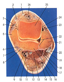
1. Tendon m. extensor hallucis longus |
6. Tendon m. tibialis -posterior |
13. Flexor hallucis longus m. |
23. Lateral anterior malleolar a. and v. and intermediate dorsal cutaneous
nerve |
This section passes through the ankle joint (talocrural articulation). The skeletal elements of the joint include the fibula (21), tibia (5, 16), and talus (25). Some important ligaments of the ankle can be identified, and these include the anterior talofibular (24), posterior tibiofibular (17), flexor retinaculum (7), and the medial (deltoid) ligament (3). The blood supply includes the anterior tibial (26), lateral anterior malleolar (23), peroneal (18), and posterior tibial (9) arteries. The intermediate dorsal cutaneous (23), lateral and medial plantar (10), and the sural (14) nerves are identified.
The talocrural articulation is a compound hinge joint (ginglymus) formed of the inferior and malleolar articular surfaces of the tibia, the malleolar articular surface of the fibula, the trochlea, and the superior surface and the medial and lateral malleolar surfaces of the talus. These bones are united by an articular capsule enclosing the joint cavity. The capsule is supplemented by the medial (deltoid), anterior talofibular, posterior talofibular, and calcaneofibular ligaments. The medial (deltoid) ligament is composed of the tibionavicular, talocalcaneal, anterior tibiotalar, and posterior tibiotalar parts.
The articular cavity (22) is very extensive. Besides following the space enclosed within the articular capsule of the ankle joint, it may extend proximally, between the tibia and fibula, as far as the interosseous membrane.
Next Page | Previous Page | Section Top | Title Page
Please send us comments by filling out our Comment Form.
Anatomy Atlases is licensed under a Creative Commons Attribution-NonCommercial-ShareAlike 4.0 International License. 
"Anatomy Atlases", the Anatomy Atlases logo, and "A digital library of anatomy information" are all Trademarks of Michael P. D'Alessandro, M.D.
Anatomy Atlases is funded in whole by Michael P. D'Alessandro, M.D. Advertising is not accepted.
Your personal information remains confidential and is not sold, leased, or given to any third party be they reliable or not.
The information contained in Anatomy Atlases is not a substitute for the medical care and advice of your physician. There may be variations in treatment that your physician may recommend based on individual facts and circumstances.
URL: http://www.anatomyatlases.org/