Atlas of Human Anatomy in Cross Section
Ronald A. Bergman, Ph.D., Adel K. Afifi, M.D., Jean J. Jew, M.D., and Paul
C. Reimann, B.S.
Peer Review Status: Externally Peer Reviewed
Sectional anatomy has had a long history, which reached a zenith in the nineteenth century only to decline in the early twentieth, and is now enjoying a renaissance because of advances in radiological techniques such as computed tomography and magnetic resonance imaging.
These newer methodologies, which in some cases approach in quality actual sections of the human body, now require that all students of anatomy-including surgeons, radiologists, anatomists, and others-be educated in sectional anatomy.
History of Sectional Anatomy
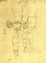
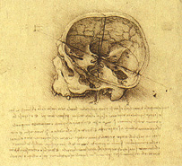
Although it cannot be stated with assurance who first began the study of sectional anatomy, it is certain that the method was in use in the early sixteenth century. Examples of anatomical sections can be found in the anatomical drawings of the Italian genius, anatomist, and artist Leonardo da Vinci (Figs. 1-5). It is also possible that Vesalius, the great Flemish anatomist, was the first to represent, in transverse sections, the brain in situ (Fig. 6).
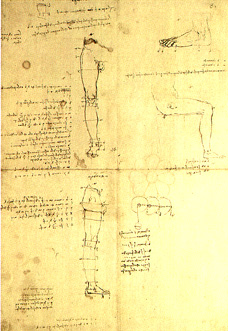 Figure 1.
|
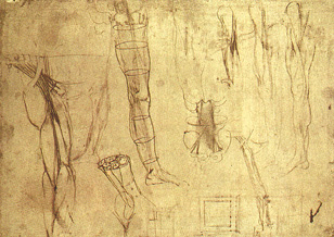 Figure 3.
|
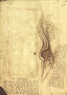 Figure 5. |
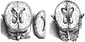 Figure 6. |
A major problem facing all these very early investigators was a lack of methods or even knowledge of existing methods to preserve and harden bodies and hold their parts in their normal position without distortion.
De Reimer, a Dutch anatomist of the early nineteenth century, gave demonstrations of transverse sections of frozen bodies. He published an atlas showing the exact position of the internal organs of the body and their relations. De Reimer stated, "Considering that the position and the condition of the internal parts of the human body can be demonstrated in a more exact manner by means of freezing than by ordinary anatomy effected with all the talent possible, I have made several sections and the results surpass all my expectations. Stimulated by the advantage that medicine and surgery may obtain from this method, which can be effected so exactly, I have resolved to give a more useful and more general exposition of this kind of work by the publication of several illustrations." De Reimer's studies of frozen bodies were entirely dependent on severe weather conditions, as no modern freezers were available to him.
In 1852, the great Russian anatomist, educator and surgeon Nicholas Pirogoff published a five-volume work entitled Anatomie Topographica, Sectionibus per corpus humanum congelatum, triplicl directione ductus illustrata. The five volumes consisted of an octavo of 786 pages of descriptive matter and four imperial folios containing 213 plates. Pirogoff, who had better climatic conditions, also used the same freezing technique employed by De Riemer, but he was unaware of the latter's priority. Assuming that he was the innovator, he states in his introduction that, "Nobody before me, as far as I know, has ever proposed or employed a method by which the human body could be cut like wood into thin sections."
In 1872, Braune published an atlas entitled Topograpisch-anatomischer Atlas nach Durchschnitten an gefrorenen Cadavern. This atlas is less extensive than Pirogoff s but is more detailed and accurate. The superiority of Braune's work reflected improvements in technique and great diligence and care in the quality of his work. Braune preserved his frozen sections by allowing them to thaw in "strong" alcohol, which was an advance introduced by Rudinger.
Rudinger published his work in 1873, and it contained 721 pages of text and 183 illustrations, 73 of which are illustrations of sections of frozen adults and newborns taken in the three principal planes. The purpose of the work, as stated in the preface, was "to present the individual structures, in various regions of the body, in comprehensive groups, thus making it of direct value in practical medicine. Not only for medicine, but also for all the phases of science and art in which anatomy takes a prominent place, does this specialized anatomical knowledge reach its true worth, both in giving a deeper insight into the topographical relations of the organs, and by enabling one to carry over from the cadaver to the living body the total picture thus obtained."
In 1881, Thomas Dwight published a small book entitled Frozen Sections of a Child. This was the first American attempt to stress the importance of studying gross anatomy using serial body sections. Dwight included 25 transverse sections through the trunk of a 3-year-old girl. Although the work was deficient in quantity and detail, it emphasized the value of this approach in learning and teaching gross anatomy.
More detailed sectional anatomical books were produced by Symington in 1887 and by Macewen in 1893. Macewen was the first to show in great detail sections of the skull and the brain in situ.
At this time, sections of frozen bodies were still not entirely satisfactory for a variety of reasons, and various methods of polishing the cut surfaces were devised. In 1909, Tunis's method consisted of "polishing sawn sections, while still frozen hard, on a rapidly revolving wooden wheel wet with water and finely powdered pumice stone." Needless to say, freezing artifacts are still a problem today for sectional anatomists and others interested in sections of the brain and other body parts.
The introduction of formalin as a tissue hardening fluid was the beginning of a new era in the study of sectional anatomy. In this regard, formalin was first introduced by Gerota (1895), who injected a 5% aqueous solution of formalin and then froze and sectioned the body.
Terry, in 1900, made an important advance when he discovered he could make excellent sections after injecting the body with a solution containing equal parts of 50% formalin and 95% alcohol without freezing. The best results, according to Terry, were obtained when the body was fixed by this solution and then thoroughly decalcified with 10% hydrochloric acid.
In 1901, Jackson reported a method that has since been used extensively. The method is simply the thorough injection of the body, via blood vessels, with a 50% aqueous solution of formalin. Jackson stated its superiority as follows: "no freezing is required; sections are made more easily and smoothly; and they do not thaw-out and become loose and flabby upon handling."
The first work of importance using formalin-hardened bodies was that done by Sellheim in 1903. His illustrations were published in a large folio consisting of 40 life-sized reproductions of sections of pelves of females of various ages, taken in the three principal planes.
Potter, in 1905, was the first to use the method of Jackson and to make an exhaustive study of the sections thus obtained. Potter published the work in a volume entitled Topography of the Thorax and Abdomen. The organs and other structures are identified in greater detail than in any previous work. Potter made use of the concept of "key figures" to advantage in this work, which otherwise suffered from plate reduction and labels that were not always clear or well placed.
In 1911, Eycleshymer and Schoemaker published their work, consisting of 113 superb tracings of sections in the transverse plane. These sections were prepared from bodies fixed in 50% formalin. The sections were traced with meticulous care. In great detail, the component parts of the sections were labeled directly on the illustration in a manner that did not unduly interfere with the examination of the illustration. This study sets the standard by which work of this kind should be measured and compared. Even with today's photographic methods it is not easy to replicate the work of Eycleshymer and Schoemaker.
Since 1911, several authors have published texts of sectional anatomy and have been successful, in varying degrees, in accomplishing their objectives. In some cases, however, the photographic quality or the material is not up to the standard set by Eycleshymer and Schoemaker.
This Atlas of Sectional Anatomy
The present work is an attempt to provide a high-quality color photographic atlas of sectional anatomy. By presenting actual photographs of sections of the human body, one of the major disadvantages of drawings, that of possible inaccuracies due to artistic interpretation, will be avoided. Moreover, high-quality photographic images offer the best means of correlation with radiological images.
The specimens used in the preparation of this atlas have come from several bodies that were embalmed by routine anatomical procedures and then thoroughly frozen. The bodies were sectioned using a bandsaw. The sections were then thawed and cleaned in a chilled solution containing 20% alcohol and 20% glycerine in distilled water. The sections were then refrozen and stored until they were photographed.
We and our colleagues at Williams & Wilkins have made every effort to try to meet the challenge and standards set by our predecessors: to provide an atlas of sections that accurately represent human anatomy and to provide present and future students of anatomy and allied fields with a useful and valued resource.
Explanatory Note
The body has been arbitrarily divided into sections. This has been a matter of convenience. Section I needs no explanation; Section 2, however, does. Because the transition between the head and thorax involves not only the neck but also the upper limb, it was thought that the most suitable subdivision of the region should include neck, shoulders, upper arm, and upper thorax. The upper thorax contains the upper half of the lungs, aortic arch, some of the great vessels, and part of the arm and the shoulder girdle. The lower thorax includes the heart and about half of the lungs. The lungs are divided, therefore, between three sections: 2, 3, and 5. This should not introduce undue difficulty in studying the lungs. The remainder of the sections are in systematic order, and whatever explanation is required for the subdivision of thorax and abdomen, abdomen and pelvis, and pelvis and lower limb is given in the annotation of the first plate in each section.
In some areas of interest, both surfaces of a section were photographed. These are included because we believe they provide additional information. Sections of the head taken "below the cranial cavity" are tilted. We used this device to show structures at two levels in the same section. Although this is not desirable, it was done to reduce cost as well as the number of sections while providing some views of cranial nerves and other structures not only adequately but well.
It is important to remember that all sections (except where noted) are viewed from below, looking toward the head. This is the way radiologists view sections of the body, unlike the usual (and more rational) anatomical view, which is looking down toward the feet.
Also importantly, the right side of the body (or section) is usually at the left side of the photograph and the left side is, therefore, usually on the right side of the photograph.
We have used several abbreviations on the labels, and these are m. for muscle, mm. for muscles, a. for artery, aa. for arteries, v. for vein, vv. for veins, br. for branch, and brs. for branches. All other words are spelled out completely.
All anatomical details included in the annotations are taken from Morris's Human Anatomy, 12th edition, B.J. Anson, ed. (published by McGraw-Hill, Blakiston Division, New York, 1966), after being checked for accuracy in current editions of both the American (30th ed.) and British (37th ed.) Gray's Anatomy. We are indebted to both the authors and publishers of these valued reference texts.
The key figures are intended to provide only an approximate positional guide to the transverse plane of each numbered section in a particular series.
Please send us comments by filling out our Comment Form.
Anatomy Atlases is licensed under a Creative Commons Attribution-NonCommercial-ShareAlike 4.0 International License. 
"Anatomy Atlases", the Anatomy Atlases logo, and "A digital library of anatomy information" are all Trademarks of Michael P. D'Alessandro, M.D.
Anatomy Atlases is funded in whole by Michael P. D'Alessandro, M.D. Advertising is not accepted.
Your personal information remains confidential and is not sold, leased, or given to any third party be they reliable or not.
The information contained in Anatomy Atlases is not a substitute for the medical care and advice of your physician. There may be variations in treatment that your physician may recommend based on individual facts and circumstances.
URL: http://www.anatomyatlases.org/