
Ronald A. Bergman, Ph.D., Adel K. Afifi, M.D., Paul M. Heidger,
Jr., Ph.D.
Peer Review Status: Externally Peer Reviewed
Most textbooks of microscopic anatomy or histology contain an abundant number of electron micrographs, and, with each new edition, they present fewer light micrographs of actual tissue sections in color. For this and other reasons, this book is devoted to illustrations of cells, tissues, and organs prepared for the light microscope.
Students of medicine, in particular, and of biology will find that the light microscope is the most important tool they will use, in spite of physical limitations in its resolving power. Advances in light microscope techniques, which use glutaraldehyde-osmium fixation, plastic embedding, and 1-µm (micron) sections, permit the highest resolution possible by this instrument.
The light microscope owes its essentiality to the vast experience gained by its use over the past 200 years. Its utility in the diagnosis of both normal and pathologic conditions is firmly established in practice. In addition, very large tissue sections may be examined (measured in centimeters), and a variety of special and selective stains may be used, enabling the study not only of cells and tissues but also of critically important large cell populations and their distribution in tissues and organs.
The electron microscope, in spite of its vastly improved resolution of exquisitely prepared tissue, is severely limited by the extremely small area of tissue (several millimeters) that can be examined, with the significant risk that important structures or structural alterations may never be seen. It is also very time-consuming to use it for surveying a significant amount of tissue adequately, and it is expensive and not widely used by practitioners of medicine and pathology except under special circumstances. Therefore, the judicious use of the light microscope is a primary prerequisite of electron microscopic study.
It is certain that future advances in electron- microscope technique will allow this instrument to perform a more important function in medical practice than it now does. Of course, its use in research on structure and function in health and disease is securely established as an important extension of the continuum of other optical systems. For the present, however, a firm background in structure, as revealed by the light microscope as well as the electron microscope, is essential to the training of the basic science researcher and to the future physician.
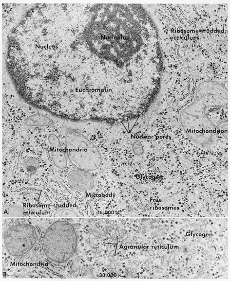
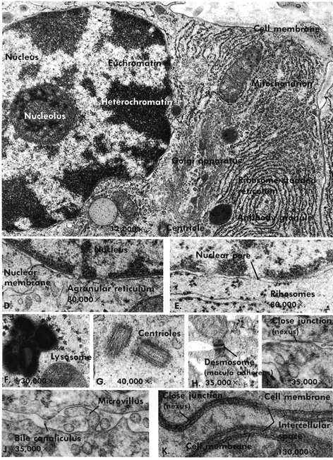
As mentioned earlier, with the exception of the accompanying plate, electron micrographs are not included in this atlas. It is believed that most textbooks of histology provide an abundant number of electron micrographs. The cellular organelles shown here by electron microscopy are cross referenced to light micrographs found elsewhere. With the exception of the lysosome, microbody, and intracellular membrane systems, every organelle and inclusion of the cell have been previously discovered by light microscopy, and their functions identified. Confirmation, details of organelle fine structure, and very important variations in intracellular and outer cell membrane ultrastructure required the resolving power of the electron microscope for their elucidation. For details, one should refer to various monographs and textbooks of histology, some of which are listed in the reference section.


|
Structure |
Primary Function |
Plate References for Corresponding Light Micrographs |
|
Nucleus |
Contains metabolically active and inactive deoxyribonucleic and ribonucleic acid |
|
|
Euchromatin |
Active deoxyribonucleic acid |
|
|
Heterochromatin |
Inactive deoxyribonucleic acid |
|
|
Nucleolus |
Ribonucleic acid, source of cytoplasmic ribonucleic acid (ribosomes) |
|
|
Nuclear pores |
Complex channels in the membrane surrounding the nuclear nucleic acid or chromatin. Important for nuclearcytoplasmic exchanges of ions, metabolites, and nuclear RNA |
-- |
|
Centriole |
Important in cell division. Modified in ciliated and sperm cells |
|
|
Ribosomes |
Free or unbound ribosomes found in cells undergoing differentiation. Synthesize protein for intracellular use |
|
|
Ribosome-studded reticulum |
Abundant in glandular tissue and nerve. Form products for extracellular digestive and other functions |
|
|
Agranular reticulum |
Important in metabolic and detoxifying functions and ion transport |
-- |
|
Golgi apparatus |
Important in concentration and packaging of secretory products such as protein, enzymes, and mucus |
|
|
Mitochondria |
The energy-producing organelle (adenosine triphosphate) and other metabolic functions |
|
|
Peroxisome (microbody) |
Contains the enzyme uricase, D-amino acid oxidase, and catalase |
-- |
|
Lysosome |
Contains hydrolytic enzymes for use in intracellular metabolic functions |
-- |
|
Antibody "granule" |
Protein antibody synthesized by the ribosome-studded reticulum, condensed into a "granule" in the Golgi apparatus |
|
|
Glycogen |
Energy-yielding metabolite in the stored form |
|
|
Cell membrane |
External limiting structure of the cells, which regulates influx and efflux of ions and metabolites. Essential in conduction of electrical impulses of muscle and nerve |
|
|
Desmosome |
Attachment sites between epithelial cells |
|
|
Close or gap junction |
Regulates intercellular interactions between epithelial cells, smooth and cardiac muscle. Site of electrical coupling of cells |
|
|
Microvilli |
Increase in absorbing surface of epithelial cells |
|
|
Bile canaliculus |
Smallest of channels formed initially of hepatic cells from liver lobule, which transport bile |
Next Page | Previous Page | Title Page
Please send us comments by filling out our Comment Form.
All contents copyright © 1995-2024 the Author(s) and Michael P. D'Alessandro, M.D. All rights reserved.
"Anatomy Atlases", the Anatomy Atlases logo, and "A digital library of anatomy information" are all Trademarks of Michael P. D'Alessandro, M.D.
Anatomy Atlases is funded in whole by Michael P. D'Alessandro, M.D. Advertising is not accepted.
Your personal information remains confidential and is not sold, leased, or given to any third party be they reliable or not.
The information contained in Anatomy Atlases is not a substitute for the medical care and advice of your physician. There may be variations in treatment that your physician may recommend based on individual facts and circumstances.
URL: http://www.anatomyatlases.org/