
Lymph node
Ronald A. Bergman, Ph.D., Adel K. Afifi, M.D., Paul M. Heidger,
Jr., Ph.D.
Peer Review Status: Externally Peer Reviewed
CELL DIVISION
Lymph node
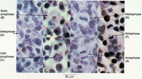
This plate illustrates the nuclear events in mitosis. They will be described in the sequence in which they occur.
Interphase (A): Non-dividing or resting stage. The chromatin appears as an irregular reticular meshwork. The nuclear membrane, or envelope, and the nucleolus are distinctly seen. Chromosomes are not visible.
Early prophase (B): Nuclear membrane and nucleolus disappear. Granularity of the nucleus is markedly increased, and filamentous structures are seen. These granules and filaments represent the chromosomes, which become shorter and thicker in this stage.
Late prophase (C): The thread- or rod-like character of the chromosomes is more apparent. Each chromosome consists of two coiled chromatids, which are not visible in this preparation. The disappearance of the nuclear membrane allows mixing of nuclear and cytoplasmic material.
Metaphase (D): Chromosomes appear condensed and line up in the equatorial plane (metaphase plate) of the cell. Each chromosome is still composed of two paired chromatids.
Anaphase (E): The daughter chromosomes (chromatids) separate and are drawn to opposite poles of the cell. They remain separate and tightly coiled, and appear at this magnification to be fused. Cytoplasmic division begins.
Telophase (F): The two distinct groups of daughter chromosomes (chromatids) appear fused and tightly packed. Cytoplasmic division is completed. Nuclear membranes re-form and nucleoli reappear.
*Helly was a twentieth-century Swiss
pathologist.
Next Page | Previous Page | Section Top | Title Page
Please send us comments by filling out our Comment Form.
All contents copyright © 1995-2024 the Author(s) and Michael P. D'Alessandro, M.D. All rights reserved.
"Anatomy Atlases", the Anatomy Atlases logo, and "A digital library of anatomy information" are all Trademarks of Michael P. D'Alessandro, M.D.
Anatomy Atlases is funded in whole by Michael P. D'Alessandro, M.D. Advertising is not accepted.
Your personal information remains confidential and is not sold, leased, or given to any third party be they reliable or not.
The information contained in Anatomy Atlases is not a substitute for the medical care and advice of your physician. There may be variations in treatment that your physician may recommend based on individual facts and circumstances.
URL: http://www.anatomyatlases.org/