
Kidney tubules medulla
Ronald A. Bergman, Ph.D., Adel K. Afifi, M.D., Paul M. Heidger,
Jr., Ph.D.
Peer Review Status: Externally Peer Reviewed
Rabbit, Helly's fluid,
iron hematoxylin-orange G, 612 x.
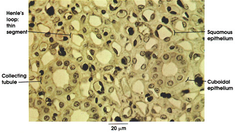
A diagnostic feature of the collecting tubules of the kidney is the appearance of distinct cell boundaries. The collecting tubules, under the influence of the antidiuretic hormone that is secreted into the vascular system in the posterior lobe of the pituitary gland (hypophysis), become permeable to water, which is reabsorbed into the vascular system, thereby concentrating the urine. in the absence of the antidiuretic hormone, the urine is dilute or hypotonic with respect to the blood.
Squamous epithelium: Single layer of flattened or squamous cells with ovoid nuclei bulging into the lumen of the thin loop of Henle*. The thin loop of Henle serves an essential role in concentrating urine, rendering it hypertonic with respect to blood plasma.
Cuboidal epithelium: Single layer of cuboidal cells (height and width of cells about equal) lining the collecting tubule. Spherical, darkly staining nuclei; clear cytoplasm.
*Henle was a nineteenth-century German anatomist, histologist, and pathologist.
Next Page | Previous Page | Section Top | Title Page
Please send us comments by filling out our Comment Form.
All contents copyright © 1995-2024 the Author(s) and Michael P. D'Alessandro, M.D. All rights reserved.
"Anatomy Atlases", the Anatomy Atlases logo, and "A digital library of anatomy information" are all Trademarks of Michael P. D'Alessandro, M.D.
Anatomy Atlases is funded in whole by Michael P. D'Alessandro, M.D. Advertising is not accepted.
Your personal information remains confidential and is not sold, leased, or given to any third party be they reliable or not.
The information contained in Anatomy Atlases is not a substitute for the medical care and advice of your physician. There may be variations in treatment that your physician may recommend based on individual facts and circumstances.
URL: http://www.anatomyatlases.org/