
Ronald A. Bergman, Ph.D., Adel K. Afifi, M.D., Paul M. Heidger,
Jr., Ph.D.
Peer Review Status: Externally Peer Reviewed
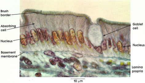
Cat, Helly's fluid, Mallory's stain, 1416 x.
Absorbing cell: Single cell layer of tall columnar cells. Basal ovoid nucleus. Although the cells appear retangular in this section, they are actually five- or six-sided when they are cross-sectioned. When the underlying tissue folds or bends, these cells may have a pyramidal appearance.
Nucleus: Ovoid. Situated in lower half of the columnar cell. The nuclei in tightly packed cells may appear elongated and staggered at different levels within the cell. This is readily seen in pseudostratified ciliated columnar (see Plate 19).
Brush border: Also know as the straited border. Made up of fine, closely packed microvilli that vastly increase the surface area of the cell. Characteristic of absorptive surfaces. Adequate absorption of digestive products is dependent upon this cell surface specialization of absorbing columnar epithelial cells.
Goblet cell: Unicellular mucous glands scattered among the tall columnar cells appear empty because mucin is extracted during tissue processing. See Plate 20 for goblet cell mucus. These unicellular gland cells are a specialization of simple epithelium and serve a protective function for the principal epithelial cell type.
Basement membrane: Delicate in appearance but a firm support for the columnar cells. See also Plate 20.
Lamina propria: Connective tissue stroma. Reticular framework containing a.variety of wandering cells as well as vascular and lymphatic channels. Cells commonly found in the lamina propria include lymphocytes, plasma cells, eosinophils, and mast cells. See also Plate 198.
Next Page | Previous Page | Section Top | Title Page
Please send us comments by filling out our Comment Form.
All contents copyright © 1995-2024 the Author(s) and Michael P. D'Alessandro, M.D. All rights reserved.
"Anatomy Atlases", the Anatomy Atlases logo, and "A digital library of anatomy information" are all Trademarks of Michael P. D'Alessandro, M.D.
Anatomy Atlases is funded in whole by Michael P. D'Alessandro, M.D. Advertising is not accepted.
Your personal information remains confidential and is not sold, leased, or given to any third party be they reliable or not.
The information contained in Anatomy Atlases is not a substitute for the medical care and advice of your physician. There may be variations in treatment that your physician may recommend based on individual facts and circumstances.
URL: http://www.anatomyatlases.org/