
Eye choroid layer
Ronald A. Bergman, Ph.D., Adel K. Afifi, M.D., Paul M. Heidger,
Jr., Ph.D.
Peer Review Status: Externally Peer Reviewed
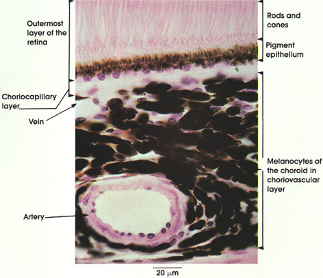
Rhesus monkey, Helly's fluid, H. & E., 612 x.
The choroid layer of the eye is a highly vascular and pigmented coat surrounding the retina. Shown in this figure is a part of the retina adjoining the choroid layer, as well as the major choroid layers. In the outermost layer of the retina, the following structures are seen:
Rods and cones: Neuroepithelial cells sensitive to light, arranged vertically and parallel. (See also Plates 306, 307, 308.)
Pigment epithelium: Single layer of pigmented cuboidal epithelial cells firmly bound to the choroid layer. Contains melanin pigment. In retinal detachments, the pigment epithelium remains attached to the choroid. The two major layers of the choroid seen in this plate are the following:
Choriocapillary layer: Composed of a network of wide lumen capillaries disposed in one plane and separated by delicate connective tissue fibers. Note that pigmented cells are essentially lacking in this layer. This layer supplies nutrition to the cells of the outermost layers of the retina.
Choriovascular layer: Filled with pigmented cells (melanin) and large-sized vessels.
Next Page | Previous Page | Section Top | Title Page
Please send us comments by filling out our Comment Form.
All contents copyright © 1995-2024 the Author(s) and Michael P. D'Alessandro, M.D. All rights reserved.
"Anatomy Atlases", the Anatomy Atlases logo, and "A digital library of anatomy information" are all Trademarks of Michael P. D'Alessandro, M.D.
Anatomy Atlases is funded in whole by Michael P. D'Alessandro, M.D. Advertising is not accepted.
Your personal information remains confidential and is not sold, leased, or given to any third party be they reliable or not.
The information contained in Anatomy Atlases is not a substitute for the medical care and advice of your physician. There may be variations in treatment that your physician may recommend based on individual facts and circumstances.
URL: http://www.anatomyatlases.org/