
Tibia knee joint
Ronald A. Bergman, Ph.D., Adel K. Afifi, M.D., Paul M. Heidger,
Jr., Ph.D.
Peer Review Status: Externally Peer Reviewed
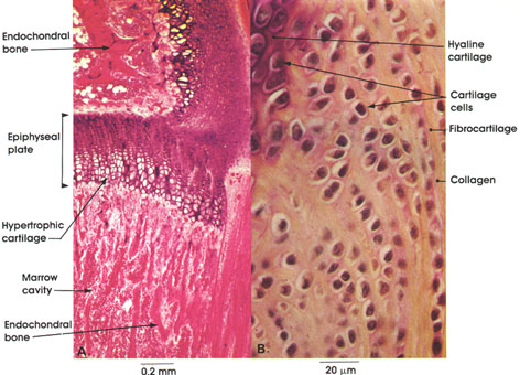
Rat, 10% formalin, A., H. & E., 50 x;
B. Gomori's aldehyde fuchsin, 612 x.
Enclochondral bone formation is a process in which an embryonal type of hyaline cartilage precedes bone formation. In A, a stage in the endochondral ossification in a long bone (tibia) is shown. Note the cartilaginous epiphyseal plate that separates the epiphysis (above) from the diaphysis (below). The epiphyseal plate is the source of new cartilage, which is replaced by bone during growth in length. Note the zone of hypertrophic cartilage within the epiphyseal plate. This is a stage in endochondral bone formation preceding calcification. Islands of formed endochondral bone are seen above and below the epiphyseal plate in the epiphysis and diaphysis. Note the marrow cavity between plates of endochrondral bone in the diaphysis. This cavity is formed by resorption of endochondral bone.
In B, note the characteristic grouping of fibrocartilage cells and their arrangement in rows separated by dense collagenous connective tissue. Adjacent to fibrocartilage, note the hyaline cartilage cells.
Next Page | Previous Page | Section Top | Title Page
Please send us comments by filling out our Comment Form.
All contents copyright © 1995-2024 the Author(s) and Michael P. D'Alessandro, M.D. All rights reserved.
"Anatomy Atlases", the Anatomy Atlases logo, and "A digital library of anatomy information" are all Trademarks of Michael P. D'Alessandro, M.D.
Anatomy Atlases is funded in whole by Michael P. D'Alessandro, M.D. Advertising is not accepted.
Your personal information remains confidential and is not sold, leased, or given to any third party be they reliable or not.
The information contained in Anatomy Atlases is not a substitute for the medical care and advice of your physician. There may be variations in treatment that your physician may recommend based on individual facts and circumstances.
URL: http://www.anatomyatlases.org/