
Ronald A. Bergman, Ph.D., Adel K. Afifi, M.D., Paul M. Heidger,
Jr., Ph.D.
Peer Review Status: Externally Peer Reviewed
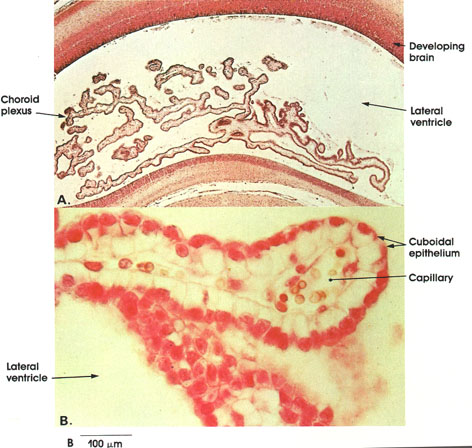
Cat, 10% formalin, H. & E., A. 8 x; B. 162 x.
A: A low-magnification plate showing the branched projections of the choroid plexus within the cavity of the lateral ventricle in a developing cat brain.
B: A high-magnification plate showing the histology of the choroid plexus. Note the single layer of cuboidal epithelium with large spherical nuclei. Beneath the epithelium is a connective tissue core containing vascular channels. The choroid plexus is a major site for production of cerebrospinal fluid.
Next Page | Previous Page | Section Top | Title Page
Please send us comments by filling out our Comment Form.
All contents copyright © 1995-2024 the Author(s) and Michael P. D'Alessandro, M.D. All rights reserved.
"Anatomy Atlases", the Anatomy Atlases logo, and "A digital library of anatomy information" are all Trademarks of Michael P. D'Alessandro, M.D.
Anatomy Atlases is funded in whole by Michael P. D'Alessandro, M.D. Advertising is not accepted.
Your personal information remains confidential and is not sold, leased, or given to any third party be they reliable or not.
The information contained in Anatomy Atlases is not a substitute for the medical care and advice of your physician. There may be variations in treatment that your physician may recommend based on individual facts and circumstances.
URL: http://www.anatomyatlases.org/