
Ronald A. Bergman, Ph.D., Adel K. Afifi, M.D., Paul M. Heidger,
Jr., Ph.D.
Peer Review Status: Externally Peer Reviewed
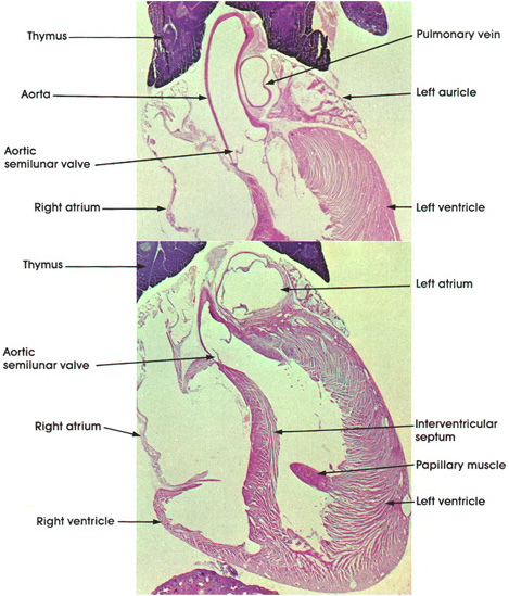
Rat, Helly's fluid, H. & E., 6 x.
This plate, in two parts, shows the four cardiac chambers and adjoining large vessels. In the upper part of the plate, the left auricle, the left ventricle, and the right atrium are seen, along with the aorta and pulmonary vein. Note the thicker muscular wall of the ventricle and the aortic semilunar valve that closes the aortic orifice. The pulmonary vein wall is thinner than the wall of the aorta. It brings oxygenated blood from the lung into the left atrium. The lower part of the plate shows both atria and ventricles as well as the aortic outlet. Note that the left ventricular wall is thicker than the right ventricular wall. This is essential for the movement of blood throughout the entire body, the systemic circulation. The interventricular septum separates the right and left ventricles and contains the conductive tissue (Purkinje fibers) of the heart. In the wall of the left ventricle note the papillary muscle. This muscle is continuous with the wall of the ventricle and projects into the cavity, where it gives origin to the chordae tendineae, which are attached to the segments of the tricuspid valve.
The thymus is seen here molded over the aortic arch. The thymus is well developed in early life and involutes after puberty. It plays a fundamental role in the development of the immune mechanism of the body.
Next Page | Previous Page | Section Top | Title Page
Please send us comments by filling out our Comment Form.
All contents copyright © 1995-2024 the Author(s) and Michael P. D'Alessandro, M.D. All rights reserved.
"Anatomy Atlases", the Anatomy Atlases logo, and "A digital library of anatomy information" are all Trademarks of Michael P. D'Alessandro, M.D.
Anatomy Atlases is funded in whole by Michael P. D'Alessandro, M.D. Advertising is not accepted.
Your personal information remains confidential and is not sold, leased, or given to any third party be they reliable or not.
The information contained in Anatomy Atlases is not a substitute for the medical care and advice of your physician. There may be variations in treatment that your physician may recommend based on individual facts and circumstances.
URL: http://www.anatomyatlases.org/