
Ronald A. Bergman, Ph.D., Adel K. Afifi, M.D., Paul M. Heidger,
Jr., Ph.D.
Peer Review Status: Externally Peer Reviewed

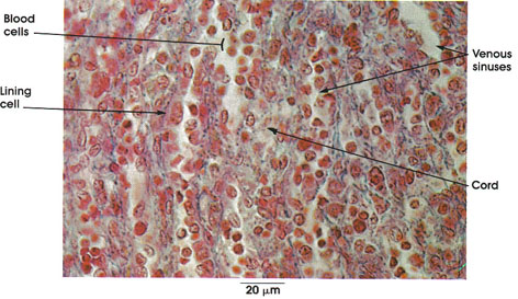
Human, Zenker's fluid, Mallory's stain, 612 x.
Venous sinuses: Form an anastomosing plexus through the red pulp between the pulp cords. Highly distensible in the living state.
Cords: Loose lymphatic tissue arranged in anastomosing cords and plates characterizes the red pulp of the spleen. Also termed Billroth or splenic cords. The cords contain varying numbers of red blood corpuscles, lymphocytes, plasma cells, and monocytes. Between cords are the venous sinusoids.
Blood cells: Fill the venous sinusoids, and impart the red color to the pulp in the fresh unfixed state.
Lining cells: Sinusoids are lined by phagocytic reticular cells belonging to the widespread macrophage (reticuloenclothelial) system. Shape changes with state of distention of the sinus.
Next Page | Previous Page | Section Top | Title Page
Please send us comments by filling out our Comment Form.
All contents copyright © 1995-2025 the Author(s) and Michael P. D'Alessandro, M.D. All rights reserved.
"Anatomy Atlases", the Anatomy Atlases logo, and "A digital library of anatomy information" are all Trademarks of Michael P. D'Alessandro, M.D.
Anatomy Atlases is funded in whole by Michael P. D'Alessandro, M.D. Advertising is not accepted.
Your personal information remains confidential and is not sold, leased, or given to any third party be they reliable or not.
The information contained in Anatomy Atlases is not a substitute for the medical care and advice of your physician. There may be variations in treatment that your physician may recommend based on individual facts and circumstances.
URL: http://www.anatomyatlases.org/