
Plate 10.190 Stomach: Fundus
Ronald A. Bergman, Ph.D., Adel K. Afifi, M.D., Paul M. Heidger,
Jr., Ph.D.
Peer Review Status: Externally Peer Reviewed
STOMACH
Fundus
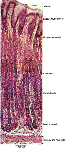
Dog, 10% formalin, H. & E., 162 x.
Although this figure is labeled fundus, it shows the characteristic glands found in most of the wall of the stomach. The term fundic as applied to this type of gland is a misnomer, since it is not limited to the fundus of the stomach but is found throughout most of the stomach wall except the cardiac and pyloric ends. Another term applied to these glands is gastric. The fundic or gastric glands are simple (sometimes slightly branched), long tubular glands that extend throughout the mucosa down to the muscularis mucosae. Note the secreted mucus covering the surface of the epithelium and the surface mucous cells with their characteristic basal nuclei and clear cytoplasm. Fundic or gastric glands contain chief and parietal cells, as well as mucus-secreting cells of the narrow neck region known as mucous neck cells. The former are the more abundant and have basally located nuclei and basophilic cytoplasm. They secrete pepsinogen. The parietal cells are larger and less abundant than the chief cells among which they are scattered. Their cytoplasm is eosinophilic and nuclei are centrally placed. They secrete HCl and, in humans, intrinsic or antipernicious anemia factor that binds and enhances the absorption of vitamin B12 by the ileum. See Plates 5 and 191.
The lamina propria is scanty and fills in the narrow spaces between glands. The muscularis mucosae is thin and arranged in layers. Delicate muscular strands extend between glands.
Next Page | Previous Page | Section Top | Title Page
Please send us comments by filling out our Comment Form.
All contents copyright © 1995-2024 the Author(s) and Michael P. D'Alessandro, M.D. All rights reserved.
"Anatomy Atlases", the Anatomy Atlases logo, and "A digital library of anatomy information" are all Trademarks of Michael P. D'Alessandro, M.D.
Anatomy Atlases is funded in whole by Michael P. D'Alessandro, M.D. Advertising is not accepted.
Your personal information remains confidential and is not sold, leased, or given to any third party be they reliable or not.
The information contained in Anatomy Atlases is not a substitute for the medical care and advice of your physician. There may be variations in treatment that your physician may recommend based on individual facts and circumstances.
URL: http://www.anatomyatlases.org/