
Plate 10.192 Duodenum and Jejunum
Ronald A. Bergman, Ph.D., Adel K. Afifi, M.D., Paul M. Heidger,
Jr., Ph.D.
Peer Review Status: Externally Peer Reviewed
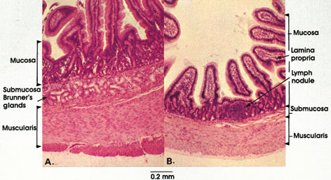
Human, 40% formalin, H. & E., 50 x.
The basic pattern and arrangement of layers in the intestinal wall are seen in both the duodenum (A) and jejunum (B). In each, there is a mucosa, submucosa, muscularis, and an adventitia or a serosa.
The mucosa has finger-like projections, the villi, lined by simple columnar epithelium. Villi of the duodenum tend to be flattened, whereas those of the jejunum are more rounded. The core of the villus is composed of loose connective tissue, blood vessels, a lymphatic vessel, smooth muscle fibers, and other cells of the connective tissue (see Plates 29, 194, and 198). This portion of the mucosa is named the lamina propria. The lamina propria terminates at the muscularis mucosae, which is composed of a band of smooth muscle fibers a few layers thick. Located within the mucosa are simple tubular intestinal glands, the crypts of Lieberkühn, and lymphatic nodules. Lymphatic nodules are found more frequently in the jejunum than in the duodenum.
The submucosa is composed of loose connective tissue and contains, in the duodenum but not the jejunum, the compound tubular mucous glands of Brunner. They are a continuation of the pyloric glands found in the stomach.
The muscularis contains an inner circular and an outer longitudinal layer of smooth muscle fibers. The two layers are separated by reticular and collagenous connective tissue containing nerve fibers and parasympathetic ganglion cells (Auerbach's plexus).
Surrounding the muscularis is the serosa, which consists primarily of loose connective tissue containing nerves, blood and lymphatic vessels, and a mesothelium. Wherever the intestine is not bound to the posterior abdominal wall, i.e., retroperitoneal, the intestine has a suspending mesentery covered with mesothelium. When the intestine is retroperitoneal, it does not have a mesentery or a mesothelial covering and the outermost layer is called adventitia. Most of the duodenum is retroperitoneal, whereas the entire jejunum is intraperitoneal.
See also Plate 199.
Next Page | Previous Page | Section Top | Title Page
Please send us comments by filling out our Comment Form.
All contents copyright © 1995-2024 the Author(s) and Michael P. D'Alessandro, M.D. All rights reserved.
"Anatomy Atlases", the Anatomy Atlases logo, and "A digital library of anatomy information" are all Trademarks of Michael P. D'Alessandro, M.D.
Anatomy Atlases is funded in whole by Michael P. D'Alessandro, M.D. Advertising is not accepted.
Your personal information remains confidential and is not sold, leased, or given to any third party be they reliable or not.
The information contained in Anatomy Atlases is not a substitute for the medical care and advice of your physician. There may be variations in treatment that your physician may recommend based on individual facts and circumstances.
URL: http://www.anatomyatlases.org/