
Plate 10.197 Duodenum
Ronald A. Bergman, Ph.D., Adel K. Afifi, M.D., Paul M. Heidger,
Jr., Ph.D.
Peer Review Status: Externally Peer Reviewed
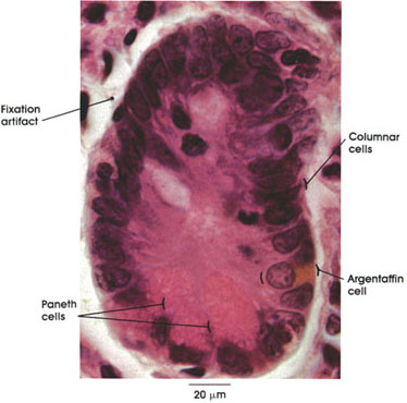
Human, 10% formalin-Zenker fixation, H. & E., 612 x.
Intestinal glands are simple tubular glands located in the mucous membrane. These glands are surrounded by a cell-rich connective tissue, the lamina propria. Intestinal glands of Lieberkühn secrete the so-called intestinal juice (succus entericus).
Columnar cells: Shorter than the columnar absorbing cells of the villi. Poorly developed striated border. Source of the surface epithelial cells at the apex of the villus.
Argentaffin cell: Also known as enterochromaffin cells. Fairly common in duodenum. Located among epithelial cells lining the crypts of Lieberkühn (intestinal glands). Contain fine granules stainable by silver salts (argentophilic) and by dichromate, and located in the abluminal portion of the cell between the nucleus and the basement membrane. Argentaffin cells are identified with the production of serotonin (5-hydroxytryptamine), which is secreted into the lamina propria rather than the intestinal lumen. Serotonin is a powerful stimulant of smooth muscle, resulting in contraction, and may play a role in stimulating peristaltic activity of the intestine.
Paneth cells: Coarsely granular cells in the depth of the intestinal gland. Acidophilic granules apically placed. The base of the cell is dark staining and basophilic. Acidophilic granules accumulate during fasting and disappear during digestion. The exact function of this cell is not established, but it has been suggested that it may secrete digestive enzymes (lipoenzyme or a peptidase, or both, and antibacterial lysozyme).
Next Page | Previous Page | Section Top | Title Page
Please send us comments by filling out our Comment Form.
All contents copyright © 1995-2024 the Author(s) and Michael P. D'Alessandro, M.D. All rights reserved.
"Anatomy Atlases", the Anatomy Atlases logo, and "A digital library of anatomy information" are all Trademarks of Michael P. D'Alessandro, M.D.
Anatomy Atlases is funded in whole by Michael P. D'Alessandro, M.D. Advertising is not accepted.
Your personal information remains confidential and is not sold, leased, or given to any third party be they reliable or not.
The information contained in Anatomy Atlases is not a substitute for the medical care and advice of your physician. There may be variations in treatment that your physician may recommend based on individual facts and circumstances.
URL: http://www.anatomyatlases.org/