
Plate 12.236 Kidney: Cortex
Ronald A. Bergman, Ph.D., Adel K. Afifi, M.D., Paul M. Heidger,
Jr., Ph.D.
Peer Review Status: Externally Peer Reviewed
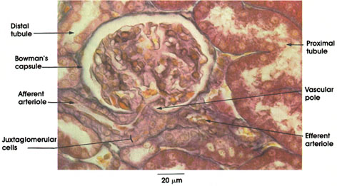
Rhesus monkey, Zenker's fluid, Mallory's stain, 612 x.
Bowman's capsule: Squamous epithelium forming a double-walled cup surrounding the glomerular capillaries. The portion of the wall applied to the capillaries is termed the visceral epithelium and can be seen in Plate 235. The visceral epithelium is separated from the outer wall by Bowman's space (Plate 234). The outer wall is continuous with the proximal tubule, and Bowman's space is continuous with the lumen of the proximal tubule at the urinary pole of the capsule.
Vascular pole: The point of entry of the afferent arteriole into Bowman's capsule. The arteriole immediately forms the tuft of glomerular capillaries and the point of origin of the efferent arteriole from glomerular capillaries, which leaves Bowman's capsule.
Afferent arteriole: Carries blood to the glomerular capillaries.
Efferent arteriole: The arterial vessel that carries blood away from the glomerular capillaries. Uniquely, in the kidney, a capillary bed is interposed between arterial vessels. The efferent arteriole leads to the cortical intertubular capillary network (see Plate 233).
Juxtaglomerular cells: Myoepithelioid cells replace typical smooth fibers in the wall of the afferent arteriole as it approaches the glomerulus. These cells secrete a hypertensive factor, renin. See also Plates 237 and 238.
Proximal tubule: Single layer of cuboidal cells with an irregular brush border.
Distal tubule: Cuboidal cells without a brush border, which stain less intensely than the proximal tubule cells.
Next Page | Previous Page | Section Top | Title Page
Please send us comments by filling out our Comment Form.
All contents copyright © 1995-2024 the Author(s) and Michael P. D'Alessandro, M.D. All rights reserved.
"Anatomy Atlases", the Anatomy Atlases logo, and "A digital library of anatomy information" are all Trademarks of Michael P. D'Alessandro, M.D.
Anatomy Atlases is funded in whole by Michael P. D'Alessandro, M.D. Advertising is not accepted.
Your personal information remains confidential and is not sold, leased, or given to any third party be they reliable or not.
The information contained in Anatomy Atlases is not a substitute for the medical care and advice of your physician. There may be variations in treatment that your physician may recommend based on individual facts and circumstances.
URL: http://www.anatomyatlases.org/