
Plate 13.249 Ovary
Ronald A. Bergman, Ph.D., Adel K. Afifi, M.D., Paul M. Heidger,
Jr., Ph.D.
Peer Review Status: Externally Peer Reviewed
Rabbit, 10% formalin, H. & E., 612 x.
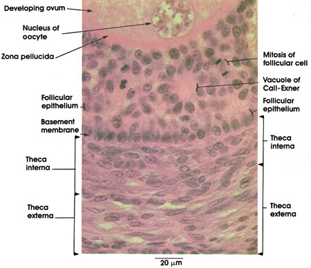
This plate shows a rather advanced stage,in the maturation of a primary ovarian follicle. Note the increase in size of the oocyte and the follicular cells in comparison to that seen in early primary follicles (Plate 246). The nucleus of the oocyte is large and has a sparse reticulated chromatin network. The cytoplasm is granular. The zona pellucida separates the oocyte from the follicular cells. This prominent layer is rich in polysaccharides and is believed to be elaborated by both oocyte and follicular cells. It persists even after the degeneration of the oocyte during atresia of the follicle (Plate 248). The follicular cells have formed a stratified epithelial layer at this stage of growth. Mitotic figures are frequent, indicating continued active proliferation. Accumulations of densely staining material are seen among follicular cells. These are vacuoles of Call-Exner*. They are believed to represent droplets within the cytoplasm of follicular cells. They stain positively with PAS and may be the precursors of follicular fluid. The stroma around the follicular epithelium is composed of an inner theca interna and an outer theca externa. A basement membrane separates the follicular cells from the theca interna; no distinct boundary exists between the thecae interna and externa. The cells of the theca interna have epithelioid characteristics and are believed to elaborate androstenedione. Note the ovoid or round nuclei of the theca interna. The theca externa is composed of spindle-shaped cells and is more fibrous. Both thecae are connective tissue derivatives.
*Call and Exner were nineteenth-century Viennese physicians and physiologists.
Next Page | Previous Page | Section Top | Title Page
Please send us comments by filling out our Comment Form.
All contents copyright © 1995-2025 the Author(s) and Michael P. D'Alessandro, M.D. All rights reserved.
"Anatomy Atlases", the Anatomy Atlases logo, and "A digital library of anatomy information" are all Trademarks of Michael P. D'Alessandro, M.D.
Anatomy Atlases is funded in whole by Michael P. D'Alessandro, M.D. Advertising is not accepted.
Your personal information remains confidential and is not sold, leased, or given to any third party be they reliable or not.
The information contained in Anatomy Atlases is not a substitute for the medical care and advice of your physician. There may be variations in treatment that your physician may recommend based on individual facts and circumstances.
URL: http://www.anatomyatlases.org/