
Plate 13.253 Mammary Gland
Ronald A. Bergman, Ph.D., Adel K. Afifi, M.D., Paul M. Heidger,
Jr., Ph.D.
Peer Review Status: Externally Peer Reviewed
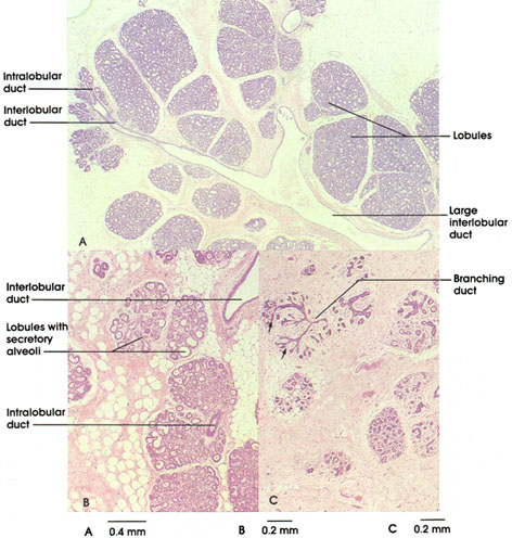
A. Human, in pregnancy, 10% formalin, 22 x.
B. Monkey, late pregnancy, 10% formalin, 36 x.
C. Human, resting, 10% formalin, 36 x.
The mammary glands are compound tubuloalveolar glands derived from the integument; they are responsive to reproductive and other hormones, and therefore undergo marked developmental, aging, and physiological changes during the course of the reproductive life of the mammalian female. Of great clinical import is the sobering statistic that cancer of the breast (usually adenocarcinoma of the ductal lining) is the most common cancer in women.
This plate illustrates distinctive differences in the histomorphology, breast tissue exhibits, depending upon physiologic state. During the development of the gland at pregnancy (A), secretory units (alveoli) bud from the ducts, and the glandular parenchyma enlarges in mass at the expense of the surrounding connective tissue. The duct system is best visualized in a gland that is "resting" (C), that is, one that is neither a gland of pregnancy nor of full lactation. Note in C how rudimentary secretory units, the alveoli (arrow), lie at the terminal arborizations of the alveolar duct system. The lobation of the gland is not well appreciated in histological section, a lobe being defined as that region of the breast drained by one lactiferous duct. The lobes are subdivided into lobules, the smallest order of which are seen here in each figure, separated by connective tissue partitions. Note the fortuitous section in A, revealing the continuity of the alveolar and higher-order system of ducts.
During pregnancy (A), the expanded alveolar component of the breast is evidenced by the highly basophilic cytoplasm of the alveolar cells, reflecting the initial synthesis of a product abundant in lactoprotein and low in lipid content (i.e., colostrum). The alveoli, however, do not reach their full degree of development until post-parturn and active lactation. Note that in the gland from late pregnancy (prelactating, B), the alveoli have expanded both in extent throughout the lobules and in size. Some alveoli are distended but exhibit regular walls and little secretory product. in contrast, the alveoli of the fully developed lactating gland typically are distended and saccular in appearance and contain a lightly eosinophilic secretory product (see Plate 254). Such alveoli are lined by a cuboidal or flattened epithelium. The alveoli and lobules are separated by sparse connective tissue, the mass of the entire gland being largely taken up with secretory alveoli and associated ducts. Like the sweat glands of the integument, the alveoli of the breast are enfolded by a network of stellate myoepithelial cells, which contract in response to oxytocin, thus facilitating the milk ejection mechanism.
The student is cautioned that the mammary gland as a whole exhibits considerable normal variation in its histological development and, in addition, undergoes cyclic changes in synchrony with the menstrual cycle. Considerable latitude is encountered even within lobules with respect to activity and distention of alveoli (e.g., B). Therefore, variation from the "ideal" description is to be expected in your laboratory study.
Next Page | Previous Page | Section Top | Title Page
Please send us comments by filling out our Comment Form.
All contents copyright © 1995-2024 the Author(s) and Michael P. D'Alessandro, M.D. All rights reserved.
"Anatomy Atlases", the Anatomy Atlases logo, and "A digital library of anatomy information" are all Trademarks of Michael P. D'Alessandro, M.D.
Anatomy Atlases is funded in whole by Michael P. D'Alessandro, M.D. Advertising is not accepted.
Your personal information remains confidential and is not sold, leased, or given to any third party be they reliable or not.
The information contained in Anatomy Atlases is not a substitute for the medical care and advice of your physician. There may be variations in treatment that your physician may recommend based on individual facts and circumstances.
URL: http://www.anatomyatlases.org/