
Plate 14.265 Seminiferous Tubule
Ronald A. Bergman, Ph.D., Adel K. Afifi, M.D., Paul M. Heidger,
Jr., Ph.D.
Peer Review Status: Externally Peer Reviewed
Rhesus monkey, Helly's fluid, iron hematoxylin and orange G stains, 612 x.
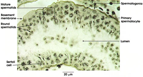
In the seminiferous tubule, spermatogenic cells are arranged in orderly layers between the basement membrane and the lumen.
Spermatogonia: Located directly above the basement membrane. Spherical nucleus. Spermatogonia are the germ cells from which spermatozoa ultimately arise. They are the only sex cells present before onset of puberty. They contain 23 pairs of chromosomes.
Primary spermatocytes: Lie in the next layer, deep to the spermatogonia. Largest germ cells. Nuclei are large and vesicular with condensed chromatin. Chromatin may appear as elongated spiremes, i.e., irregularly disposed chromatin filaments. Primary spermatocytes divide by meiosis. Meiosis is nuclear division in which the diploid chromosome number (23 pairs) is halved to the haploid number (23 single set) in the formation of sex cells.
Round spermatids: Adjacent to the lumen. Small in size. Spermatids constitute the last stage in the transformation to spermatozoa.
Mature spermatids: Heads are located near Sertoli cells, and tails project into the lumen. Heads are transformed nuclei of round spermatids. Will be released into lumen as spermatozoa.
Sertoli cells: Supporting cells of the testicular epithelium, which were first described by the Italian physiologist Enrico Sertoli in 1865. Tall columnar cells extend from the basement membrane to the lumen. Nucleus ovoid in shape with prominent nucleolus. Cell borders are difficult to outline in this preparation.
The process of spermatogenesis from spermatogonia to mature spermatozoa requires about 64 days in man.
Next Page | Previous Page | Section Top | Title Page
Please send us comments by filling out our Comment Form.
All contents copyright © 1995-2025 the Author(s) and Michael P. D'Alessandro, M.D. All rights reserved.
"Anatomy Atlases", the Anatomy Atlases logo, and "A digital library of anatomy information" are all Trademarks of Michael P. D'Alessandro, M.D.
Anatomy Atlases is funded in whole by Michael P. D'Alessandro, M.D. Advertising is not accepted.
Your personal information remains confidential and is not sold, leased, or given to any third party be they reliable or not.
The information contained in Anatomy Atlases is not a substitute for the medical care and advice of your physician. There may be variations in treatment that your physician may recommend based on individual facts and circumstances.
URL: http://www.anatomyatlases.org/