
Plate 15.286 Thyroid Gland
Ronald A. Bergman, Ph.D., Adel K. Afifi, M.D., Paul M. Heidger,
Jr., Ph.D.
Peer Review Status: Externally Peer Reviewed
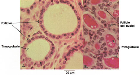
Human, 10% formalin,
A. H. & E.; B. periodic acid-Schiff and hematoxylin stains, 612
x.
Follicles: Structural units of the thyroid gland supported and separated by connective tissue. Note variation in size. A single layer of cells forms the follicle. Shape of cells reflects functional activity. Cells in these follicles are cuboidal with central, rounded nuclei, indicating normal activity.
In A, the colloid in the lumen of the follicle is not stained. In B, the colloid is specifically stained red with the periodic acid-Schiff method because of the chemical composition of colloid, which is a glycoprotein-iodine complex (thyroglobulin).
Next Page | Previous Page | Section Top | Title Page
Please send us comments by filling out our Comment Form.
All contents copyright © 1995-2024 the Author(s) and Michael P. D'Alessandro, M.D. All rights reserved.
"Anatomy Atlases", the Anatomy Atlases logo, and "A digital library of anatomy information" are all Trademarks of Michael P. D'Alessandro, M.D.
Anatomy Atlases is funded in whole by Michael P. D'Alessandro, M.D. Advertising is not accepted.
Your personal information remains confidential and is not sold, leased, or given to any third party be they reliable or not.
The information contained in Anatomy Atlases is not a substitute for the medical care and advice of your physician. There may be variations in treatment that your physician may recommend based on individual facts and circumstances.
URL: http://www.anatomyatlases.org/