
Plate 15.288 Parathyroid Gland
Ronald A. Bergman, Ph.D., Adel K. Afifi, M.D., Paul M. Heidger,
Jr., Ph.D.
Peer Review Status: Externally Peer Reviewed
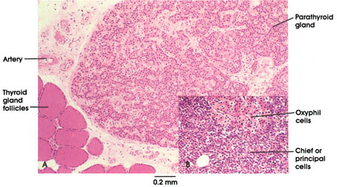
A. Rhesus monkey, glutaraldehyde, H. & E., 47 x;
B. Human, 10% formalin, H. & E., 47 x.
Two cell types are found in the parathyroid gland. The most abundant type is the chief (or principal) cell, which secretes parathyroid hormone. Chief cells have a prominent nucleus, and a cytoplasm that stains variably and may be light or dark depending upon its secretory activity.
The second type, oxyphilic (acidophilic or eosinophilic) cells, occurs in small clumps and in fewer numbers. These cells usually have small densely staining heterochromatin and an oxyphilic cytoplasm whose perimeter is usually well defined. Oxyphilic cells usually increase in number with age but their specific function is unknown.
The parathyroids are essential for life. They control blood calcium and phosphate levels. A significant decrease in blood calcium results in tetany, abnormal twitching, and muscle spasms, caused by changes in excitability at the neuromuscular junction, and death.
Dietary addition of calcium and especially administration of parathyroid hormone relieves the abnormal spasms, preventing death of the organism.
Parathyroid hormone secretion is apparently regulated by blood calcium levels only in hypoparathyroidism, phosphate levels increase and calcium levels decrease. in hyperparathyroidism, blood phosphate is low and blood calcium levels are increased. Abnormal levels of calcium may result in abnormal deposition of calcium in the kidneys and muscle. Abnormally increased blood levels of calcium occur at the expense of bone, which may fracture as a result. Calcium removal from bone is related to osteoclastic activity, which is the site of action of parathyroid hormone.
The small parathyroid glands (4 to 5 mm in diameter) vary in number from three to six and are usually found on the posterior surface of the thyroid gland.
In the lower left corner, a few follicles of the thyroid gland can be seen.
Next Page | Previous Page | Section Top | Title Page
Please send us comments by filling out our Comment Form.
All contents copyright © 1995-2025 the Author(s) and Michael P. D'Alessandro, M.D. All rights reserved.
"Anatomy Atlases", the Anatomy Atlases logo, and "A digital library of anatomy information" are all Trademarks of Michael P. D'Alessandro, M.D.
Anatomy Atlases is funded in whole by Michael P. D'Alessandro, M.D. Advertising is not accepted.
Your personal information remains confidential and is not sold, leased, or given to any third party be they reliable or not.
The information contained in Anatomy Atlases is not a substitute for the medical care and advice of your physician. There may be variations in treatment that your physician may recommend based on individual facts and circumstances.
URL: http://www.anatomyatlases.org/