
Plate 16.301 Cornea
Ronald A. Bergman, Ph.D., Adel K. Afifi, M.D., Paul M. Heidger,
Jr., Ph.D.
Peer Review Status: Externally Peer Reviewed
Rhesus monkey, 10% formalin, H. & E., 612 x.
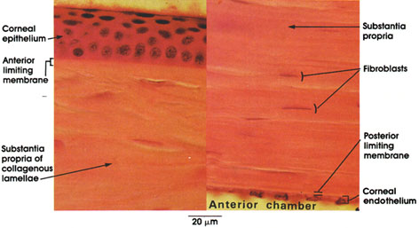
The cornea is the bulging front portion of the eye. It is nonvascular and transparent. Microscopically, five distinct layers can be recognized.
Corneal epithelium: Non-keratinized stratified squamous type of epithelium. Note that the basal layer of cells is columnar, and the most superficial are flattened.
Anterior limiting membrane: The second layer of the cornea was described by Sir William Bowman, an English surgeon, and therefore is called Bowman's membrane. This membrane appears homogeneous and structureless by light microscopy. By electron microscopy, it is shown to be composed of fine collagenous fibrils.
Substantia propria: Constitutes nine tenths of the thickness of the cornea. Composed of collagen fibrils, fibroblasts, and cementing substance. The fibrils are arranged in lamellae that run parallel to the surface of the cornea. The fibroblasts are flattened and lie between the fibrous lamellae. A mucopolysaccharicle cements the different lamellae and the collagenous fibrils within lamellae together. The metachromatic protein polysaccharide ground substance and the arrangement of fibrils within the substantia propria contribute to the transparency of the cornea.
Posterior limiting membrane: The posterior limiting membrane of the cornea was described by the French surgeon Descemet in 1758 and is known by his name. English anatomists state that it was first described by Benedict Duddell, an English oculist. This membrane appears homogeneous in the light microscope. Electron microscopy reveals a wide basement membrane made of atypical collagen.
Corneal endothelium: Low cuboidal epithelium. The term endothelium is a misnomer, since this epithelium is bathed by aqueous humor of the anterior chamber and not blood or lymph.
Next Page | Previous Page | Section Top | Title Page
Please send us comments by filling out our Comment Form.
All contents copyright © 1995-2024 the Author(s) and Michael P. D'Alessandro, M.D. All rights reserved.
"Anatomy Atlases", the Anatomy Atlases logo, and "A digital library of anatomy information" are all Trademarks of Michael P. D'Alessandro, M.D.
Anatomy Atlases is funded in whole by Michael P. D'Alessandro, M.D. Advertising is not accepted.
Your personal information remains confidential and is not sold, leased, or given to any third party be they reliable or not.
The information contained in Anatomy Atlases is not a substitute for the medical care and advice of your physician. There may be variations in treatment that your physician may recommend based on individual facts and circumstances.
URL: http://www.anatomyatlases.org/