
Plate 16.303 Ciliary Muscle and Ciliary Processes
Ronald A. Bergman, Ph.D., Adel K. Afifi, M.D., Paul M. Heidger,
Jr., Ph.D.
Peer Review Status: Externally Peer Reviewed
Rhesus monkey, 10% formalin, H. & E., 612 x.
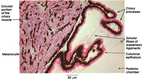
Ciliary processes: Ridges of the ciliary body as it approaches the iris run in a meridional plane. They provide an anchor for the suspensory ligaments of the lens.
Zonular fibers of suspensory ligament: Seen attached to the ciliary processes. Inelastic and radially arranged fibers from which the lens is suspended. Extend from ciliary processes to the lens capsule. When the eye is at rest, zonular fibers under tension from elastic fibers in the choroid stretch the lens. Tension in zonular fibers is reduced when ciliary muscles contract. This results in a change in the shape of the lens (accommodation). The lens becomes more spherical owing to its inherent elasticity.
Columnar epithelium: Columnar or cuboidal epithelium, which covers the ciliary processes. Indistinct cell borders. Elaborates aqueous humor.
Posterior chamber: The space between the iris and suspensory ligament of the lens. Contains aqueous humor.
Ciliary muscle: Smooth muscle fibers intermixed with melanocytes. Runs in three directions: circular, radial, and meridional. Circular fibers lie at the inner edge of the ciliary body. Contraction of ciliary muscles releases tension in suspensory ligament of the lens and of the lens capsule, thus allowing the lens to change shape to accommodate for near vision. The ciliary muscle is a continuation of the suprachoroid layer.
Melanocytes: Pigment-laden cells scattered in the connective tissue elements between muscle fibers.
Next Page | Previous Page | Section Top | Title Page
Please send us comments by filling out our Comment Form.
All contents copyright © 1995-2024 the Author(s) and Michael P. D'Alessandro, M.D. All rights reserved.
"Anatomy Atlases", the Anatomy Atlases logo, and "A digital library of anatomy information" are all Trademarks of Michael P. D'Alessandro, M.D.
Anatomy Atlases is funded in whole by Michael P. D'Alessandro, M.D. Advertising is not accepted.
Your personal information remains confidential and is not sold, leased, or given to any third party be they reliable or not.
The information contained in Anatomy Atlases is not a substitute for the medical care and advice of your physician. There may be variations in treatment that your physician may recommend based on individual facts and circumstances.
URL: http://www.anatomyatlases.org/