
Plate 16.309 Eyelid
Ronald A. Bergman, Ph.D., Adel K. Afifi, M.D., Paul M. Heidger,
Jr., Ph.D.
Peer Review Status: Externally Peer Reviewed
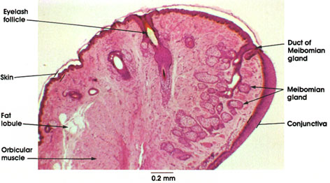
Rhesus monkey, 10% formalin, H. & E., 50 x.
The eyelid is a fold of skin; superficially, the keratinized epidermis blends internally with a mucous membrane (the conjunctiva). These layers are supported by a dermal core of connective tissue in which striated and smooth muscle, glands, and hair follicles are located.
Eyelash follicle: A row of short stout hairs are found at the free margin of the lid. Penetrate deep into the dermis. Their follicles are similar to those found elsewhere in the body but lack the arrector pili smooth muscle.
Skin: Thin layer of epidermis continuous with the conjunctiva.
Fat lobule: Scattered in the connective tissue core of the eyelid.
Orbicular muscle: Skeletal muscle bundles that lower the eyelids.
Conjunctiva: Mucous membrane lining the inside of the eyelid. The epithelium is stratified columnar with goblet cells scattered among the superficial cells.
Meibomian gland: The tarsal glands of the eyelids, first noted by Casserius in 1609 and described by Heinrich Meibom, a German anatomist, in 1666. These are simple, branched alveolar sebaceous glands disposed in a plane perpendicular to the lid margin. The glandular alveoli are connected by short lateral ducts to a long central excretory duct lined with stratified squamous epithelium. The glands open at the inner free margin of the lid at the junction of the skin and conjunctiva. Secretion of the glands serves to lubricate the surface of the lids.
Next Page | Previous Page | Section Top | Title Page
Please send us comments by filling out our Comment Form.
All contents copyright © 1995-2024 the Author(s) and Michael P. D'Alessandro, M.D. All rights reserved.
"Anatomy Atlases", the Anatomy Atlases logo, and "A digital library of anatomy information" are all Trademarks of Michael P. D'Alessandro, M.D.
Anatomy Atlases is funded in whole by Michael P. D'Alessandro, M.D. Advertising is not accepted.
Your personal information remains confidential and is not sold, leased, or given to any third party be they reliable or not.
The information contained in Anatomy Atlases is not a substitute for the medical care and advice of your physician. There may be variations in treatment that your physician may recommend based on individual facts and circumstances.
URL: http://www.anatomyatlases.org/