
Plate 16.311 Organ of Corti
Ronald A. Bergman, Ph.D., Adel K. Afifi, M.D., Paul M. Heidger,
Jr., Ph.D.
Peer Review Status: Externally Peer Reviewed
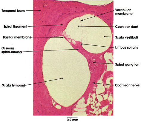
Cat, Müller's* fluid, H. & E., 50 x.
Temporal bone: The bone in which the cochlea is located.
Spiral ligament: A projection of thickened periosteum along the outer wall of the osseous canal of the cochlea.
Osseous spiral lamina: A bony shelf projecting from the modiolus across the osseous canal of the cochlea. Follows the spiral turns of the cochlea. Divides the osseous canal of the cochlea into an upper scala vestibuli and a lower scala tympani. The former is continuous with the oval window; the latter ends at the round window. The two scalae are continuous at the helicotrema.
Vestibular membrane: Also called Reissner's* membrane. A thin membrane extending from the upper surface of the limbus spiralis to the upper part of the spiral ligament. It forms the roof of the cochlear duct, which is a part of the membranous labyrinth and houses the organ of hearing.
Limbus spiralis: A thickening of periosteal connective tissue at the outer border of the osseous spiral lamina. The vestibular membrane attaches to the upper surface of the limbus.
Basilar membrane: Forms the base of the cochlear duct. Gives support to the organ of Corti.
Spiral ganglion: Ganglion of the bipolar cells. Peripheral processes contact the hair cells of the organ of Corti, and the longer central processes form the cochlear nerve.
Cochlear nerve: Formed by the central processes of the bipolar cells of the spiral ganglion, these nerve fibers, upon entering the brain stem, terminate in the dorsal and ventral cochlear nuclei (see Plates 105 and 332).
*Müller and Reissner were nineteenth-century German anatomists.
Next Page | Previous Page | Section Top | Title Page
Please send us comments by filling out our Comment Form.
All contents copyright © 1995-2025 the Author(s) and Michael P. D'Alessandro, M.D. All rights reserved.
"Anatomy Atlases", the Anatomy Atlases logo, and "A digital library of anatomy information" are all Trademarks of Michael P. D'Alessandro, M.D.
Anatomy Atlases is funded in whole by Michael P. D'Alessandro, M.D. Advertising is not accepted.
Your personal information remains confidential and is not sold, leased, or given to any third party be they reliable or not.
The information contained in Anatomy Atlases is not a substitute for the medical care and advice of your physician. There may be variations in treatment that your physician may recommend based on individual facts and circumstances.
URL: http://www.anatomyatlases.org/