
Plate 17.317 Spinal Cord
Ronald A. Bergman, Ph.D., Adel K. Afifi, M.D., Paul M. Heidger,
Jr., Ph.D.
Peer Review Status: Externally Peer Reviewed
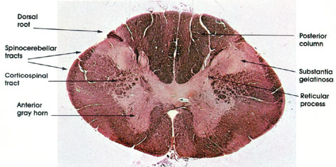
Human, 10% formalin, Weigert-carmine, 11 x.
Posterior column: The white matter located between the posterior median septum and the medial border of the posterior horn. Contains heavily myelinated nerve fibers that form the ascending gracile and cuneate tracts. These tracts carry impulses from proprioceptive and tactile receptors (Pacinian corpuscles, Golgi tendon organs, neuromuscular spindles, and Meissner's corpuscles), which give rise to sensations of position, movement, and tactile localization. These receptors are seen in Plates 120, 122, and 124.
Substantia gelatinosa: Appears as a cap-like structure at the head of the posterior horn. It is largest in the first cervical and in the lumbosacral segments but extends the whole length of the cord. This nucleus contains small cells about 6 to 20 µm in diameter and is the primary associative center of the posterior horn for incoming impulses carried by the dorsal root. This nucleus is an important part of the pathway for pain, temperature, and some tactile impulses.
Dorsal root: Sensory nerve fibers entering the spinal cord. Follow some of these fibers to the anterior horn where they terminate on motor neurons.
Spinocerebellar tracts: The spinocerebellar tracts convey impulses to the cerebellum from muscle, tendon, and joint proprioceptors, thus enabling the cerebellum to coordinate skeletal muscle activity (posture and movement).
Reticular process: Contains small and medium-sized cells, which send their axons to the adjacent as well as to the opposite anterolateral white column. Also known as the nucleus reticularis.
Anterior gray horn: Contains cells whose axons pass to the extrafusal fibers of striated skeletal muscle. The cells are the largest found in the spinal cord. These multipolar neurons have as many as 20 dendrites, and axons approximately 12 µm in diameter. Smaller neurons with thin axons (gamma efferent fibers) supply the intrafusal muscle fibers of the neuromuscular spindle (see Plates 120 and 121).
Next Page | Previous Page | Section Top | Title Page
Please send us comments by filling out our Comment Form.
All contents copyright © 1995-2024 the Author(s) and Michael P. D'Alessandro, M.D. All rights reserved.
"Anatomy Atlases", the Anatomy Atlases logo, and "A digital library of anatomy information" are all Trademarks of Michael P. D'Alessandro, M.D.
Anatomy Atlases is funded in whole by Michael P. D'Alessandro, M.D. Advertising is not accepted.
Your personal information remains confidential and is not sold, leased, or given to any third party be they reliable or not.
The information contained in Anatomy Atlases is not a substitute for the medical care and advice of your physician. There may be variations in treatment that your physician may recommend based on individual facts and circumstances.
URL: http://www.anatomyatlases.org/