
Plate 17.328 Medulla Oblongata
Ronald A. Bergman, Ph.D., Adel K. Afifi, M.D., Paul M. Heidger,
Jr., Ph.D.
Peer Review Status: Externally Peer Reviewed
Human, 10% formalin, Pal-Weigert and carmine stains, 7 x.
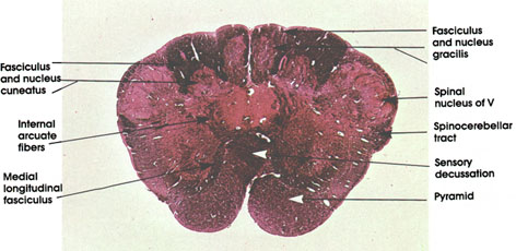
Fasciculus and nucleus gracilis: Note the reduction in size of the fasciculus gracilis as the nucleus gracilis develops. Fibers of the fasciculus synapse on neurons of the nucleus gracilis. The nucleus gracilis appears caudal to the appearance of and terminates caudal to the termination of nucleus cuneatus (see Plates 324 and 329).
Fasciculus and nucleus cuneatus: Note that the nucleus cuneatus is not as well developed at this level as the nucleus gracilis. The fasciculus cuneatus is voluminous.
Internal arcuate fibers: Second-order fibers arise from gracile and cuneate nuclei, course in the tegmentum of the medulla, and cross in the sensory decussation to form the medial lemniscus. They convey the same modalities of sensation as the gracile and cuneate tracts (proprioception, touch, and vibratory sense).
Medial longitudinal fasciculus: Descending portion of a fiber system with ascending and descending components. Arises from various brain stem nuclei, but with a major vestibular component. This system is concerned with eye and neck movements. The fibers in it are destined to synapse on the motor neurons in the cervical region supplying neck musculature.
Spinal nucleus of V: Continuation of same nucleus seen at more caudal levels (see Plate 324).
Spinocerebellar tract: Continuation of the sme tract seen at several caudal levels (see Plates 317, 324 and 327).
Sensory decussation: Also known as decussation of the medial lemniscus. Internal arcuate fibers cross here to form the contralateral medial lemniscus. Provides an anatomical basis for sensory representation of one half of the body in the contralateral cerebral cortex.
Pyramid: Same system as described in Plate 324. Descending corticospinal fibers. Lesion will result in contralateral weakness or paralysis of the upper motor neuron variety.
Next Page | Previous Page | Section Top | Title Page
Please send us comments by filling out our Comment Form.
All contents copyright © 1995-2024 the Author(s) and Michael P. D'Alessandro, M.D. All rights reserved.
"Anatomy Atlases", the Anatomy Atlases logo, and "A digital library of anatomy information" are all Trademarks of Michael P. D'Alessandro, M.D.
Anatomy Atlases is funded in whole by Michael P. D'Alessandro, M.D. Advertising is not accepted.
Your personal information remains confidential and is not sold, leased, or given to any third party be they reliable or not.
The information contained in Anatomy Atlases is not a substitute for the medical care and advice of your physician. There may be variations in treatment that your physician may recommend based on individual facts and circumstances.
URL: http://www.anatomyatlases.org/