
Plate 17.331 Medulla Oblongata
Ronald A. Bergman, Ph.D., Adel K. Afifi, M.D., Paul M. Heidger,
Jr., Ph.D.
Peer Review Status: Externally Peer Reviewed
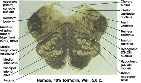
Choroid plexus: Located in the caudal part of the roof of the fourth ventricle.
Inferior vestibular nucleus: One of four vestibular nuclei. Characteristically located medial and dorsal to the restiform body, and traversed by myelinated bundles.
Tractus solitarius: Contains general visceral as well as special visceral (taste) fibers from the vagus, glossopharyngeal, and facial nerves. Fibers project onto neurons in the nucleus solitarius located in close proximity to the tract.
Nucleus solitarius: Located in close proximity to the tractus solitarius, from which it receives fibers.
Hypoglossal (CN XII) nucleus: A group of large neurons located dorsal to the medial longitudinal fasciculus in the floor of the fourth ventricle in a paramedian position. Rootlets of hypoglossal nerve course in tegmentum of medulla between the medial lemniscus and inferior olive.
Hypoglossal (CN XII) rootlets: Coursing in the tegmentum of the medulla oblongata between the medial lemniscus and the inferior olive. Exit from the ventral surface of the medulla between the pyramid and inferior olive.
Dorsal accessory olive: A component of the inferior olivary complex located dorsal to the principal olive.
Medial longitudinal fasciculus: Descending portion of a fiber system with ascending and descending components. Neurons of origin are from various brain stem nuclei, but with a major vestibular component. The fibers descending in this fasciculus are destined to synapse on motor neurons in the cervical region of the spinal cord, which supply neck musculature.
Accessory cuneate nucleus: Receives fibers of the dorsal spinocerebellar tract entering the spinal cord above the eighth cervical segment. Projects to the cerebellum via the restiform body.
Restiform body: Also known as the inferior cerebellar peduncle. A compact bundle of nerve fibers connecting the medulla with the cerebellum. Described first in 1695 and named by Humphrey Ridley, an English anatomist. Tracts and fibers forming this bundle originate in the medulla and the spinal cord.
Nucleus of spinal tract of trigeminal (CN V) nerve: Receives exteroceptive fibers from the ipsilateral side of the face via the spinal tract of the trigeminal nerve. Lesions result in loss of pain sensation in the ipsilateral face.
Medial lemniscus: Axons of gracile and cuneate nuclei. Forward continuation of the same structure seen in more caudal sections.
Principal inferior olive: Located dorsal and lateral to the pyramid. Note the characteristic convoluted appearance. The principal inferior olive is the largest component of the inferior olivary complex, which includes, in addition, the dorsal accessory inferior olive and the medial accessory inferior olive.
Pyramid: Heavily myelinated motor fiber system. Represents descending fibers from the cerebral cortex that pass through the internal capsule, cerebral peduncle, and pons before reaching the medullary pyramids. Fibers in the pyramid undergo partial crossing in the motor decussation to give rise to the lateral corticospinal tract. It is estimated that, in man, about one million fibers are present in each pyramid.
Next Page | Previous Page | Section Top | Title Page
Please send us comments by filling out our Comment Form.
All contents copyright © 1995-2025 the Author(s) and Michael P. D'Alessandro, M.D. All rights reserved.
"Anatomy Atlases", the Anatomy Atlases logo, and "A digital library of anatomy information" are all Trademarks of Michael P. D'Alessandro, M.D.
Anatomy Atlases is funded in whole by Michael P. D'Alessandro, M.D. Advertising is not accepted.
Your personal information remains confidential and is not sold, leased, or given to any third party be they reliable or not.
The information contained in Anatomy Atlases is not a substitute for the medical care and advice of your physician. There may be variations in treatment that your physician may recommend based on individual facts and circumstances.
URL: http://www.anatomyatlases.org/